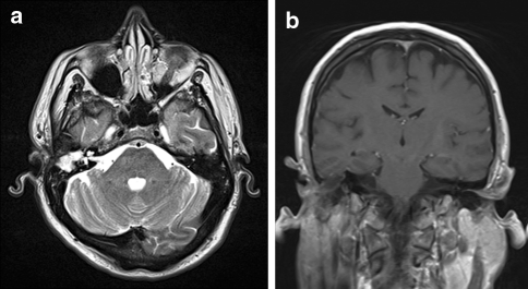
Fig. 2.
Axial T2-weighted MRI (a) on a 1.5 T MRI demonstrating a soft tissue mass originating in the right external auditory canal and middle ear with dehiscence of the right tegmen tympani. Coronal T1-weighted MRI (b) showing extension superiorly into the right middle cranial fossa where the mass abuts the dura at the inferior aspect of the right inferolateral temporal lobe

