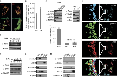
Figure 1.
Trichoplein/mitostatin is enriched at the endoplasmic reticulum–mitochondria interface. (A) Upper panels: representative confocal images of HeLa cells co-transfected with TpMs–GFP (green) and mtRFP (mito, red) or erRFP (ER, red). Lower panels: confocal images of HeLa cells co-transfected with TpMs–V5 and empty vector (left) or YFP-LC3 (autophago, right), fixed after 24 h and immunostained with TRITC-conjugated anti-V5 (red) and with an FITC-conjugated cM6PR antibody (endosomes, green) in the left panel. Scale bar, 20 μm. (B) Mean±s.e. (n=3) of interaction data from (A). (C) Left: crude HeLa mitochondria (5 mg/ml) were incubated where indicated with proteinase K (100 μg/ml). Mitochondria were in isolation buffer (Frezza et al, 2007) or in 20 mM HEPES (pH 7.4; swelling) or in 0.1% Triton X-100. Proteins (25 μg) separated by SDS–PAGE were immunoblotted with the indicated antibodies. Right: crude HeLa mitochondria were incubated in 0.1 M Na2CO3 (pH 11.3; 30 min; 4°C). After centrifugation, proteins (25 μg) from pellet (p) and supernatant (sn) separated by SDS–PAGE were immunoblotted with the indicated antibodies. (D) Real-time PCR of plectin 1b levels from HeLa cells transfected with the indicated siRNA. (E) Proteins (20 μg) from HeLa cells transfected as indicated were separated by SDS–PAGE and immunoblotted using the indicated antibodies. (F) Mitochondria were isolated at indicated times from 5 × 10⁸ HeLa cells transfected as indicated and proteins (25 μg) were separated by SDS–PAGE and immunoblotted. (G,H) Proteins (40 μg) from Percoll-purified subcellular fractions of mouse liver, were separated by SDS–PAGE and immunoblotted. (I) Representative confocal images of HeLa cells co-transfected with the indicated plasmids. After 24 h cells were fixed and immunostained with FITC-conjugated V5 antibody. Boxed areas are magnified × 9. Scale bar, 20 μm. White in the merge image: overlap of the three channels. Cyto, cytosol; ER, endoplasmic reticulum; ER-RFP, ER-targeted red fluorescent protein; FITC, fluorescein isothiocyanate; GFP, green fluorescent protein; GRP75, glucose regulatory protein 75; LC3, microtuble-associated protein 1 light chain 3; LM, light membranes; MAM, mitochondria-associated membrane; mito, mitochondria; mtRFP, matrix red fluorescent protein; SDS–PAGE, sodium dodecyl sulphate–polyacrylamide gel electrophoresis; scr, scrambled; siRNA, small interfering RNA; TpMs, trichoplein/mitostatin; TRITC, tetramethyl rhodamine iso-thiocyanate; YFP, yellow fluorescent protein.

