Figure 2.
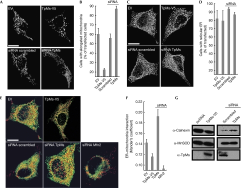
Trichoplein/mitostatin regulates mitochondrial shape and mitochondria–endoplasmic reticulum juxtaposition. (A) Representative confocal images of HeLa cells co-transfected with mtRFP and the indicated plasmids. Scale bar, 20 μm. (B) Mean±s.e.m. (n=3) of morphometric analysis from (A). (C) Representative three-dimensional reconstructions of ER HeLa cells co-transfected with ER-YFP and the indicated plasmids. Scale bar, 20 μm. (D) Mean±s.e.m. (n=3) of morphometric analysis from (C). (E) Representative three-dimensional reconstructions of ER and mitochondria in HeLa cells co-transfected with mtRFP, ER-YFP and the indicated plasmids. Yellow, organelles are closer than around 270 nm. Scale bar, 20 μm. (F) Mean±s.e. (n=5) of interaction data from (E). (G) A total of 25 μg of proteins from mitochondria isolated after 24 (left) or 48 h (right) from HeLa cells (5 × 10⁸), transfected as indicated, were analysed by SDS–PAGE and immunoblotting. ER, endoplasmic reticulum; ER-RFP, ER-targeted red fluorescent protein; Mfn2, mitofusin 2; MtRFP, matrix red fluorescent protein; SDS–PAGE, sodium dodecyl sulphate–polyacrylamide gel electrophoresis; siRNA, small interfering RNA; TpMs, trichoplein/mitostatin; YFP, yellow fluorescent protein.

