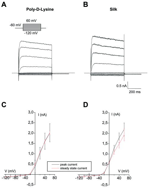
Figure 4.
Representative current traces evoked in astrocytes with a family of voltage steps from Vh of -60 mV, from -120 to 60 mV in 20 mV increments (inset). Typical currents depicting K+ current are evoked in poly-D-lysine astrocytes (A) and silk-treated astrocytes (B) at potential more positive than -40 mV. The currents had an instantaneous activation and did not display time-dependent inactivation in either experimental condition. C-D) I-V plot: Mean of current intensity recorded at peak (black lines) and steady state (red lines) in poly-D-lysine astrocytes (C) and silk-plated astrocytes (D). The outward potassium current has voltage- and time-dependence comparable in both experimental conditions and resembles those of delayed rectifier potassium channels. (n=4 for poly-D-Lysine coated and n=4 for silk).

