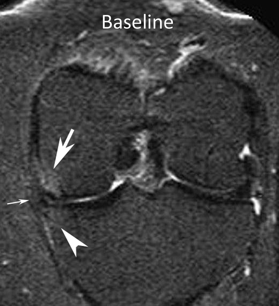
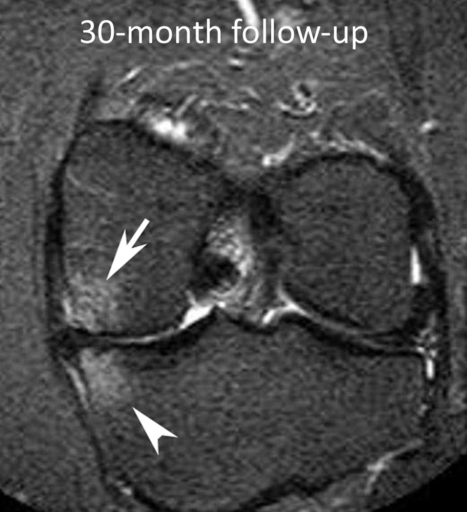
Figure 3.
Baseline: Mid coronal STIR MRI shows extrusion of the partially macerated body of the medial meniscus (small arrow) with small (WORMS grade 1) bone marrow lesions (BMLs) of the central medial femur (arrow) and tibia (arrowhead). 30-month follow-up: Coronal STIR MRI at the same level 30 months later shows enlarging BMLs (WORMS grade 2) of the central medial femur (arrow) and tibia (arrowhead).


