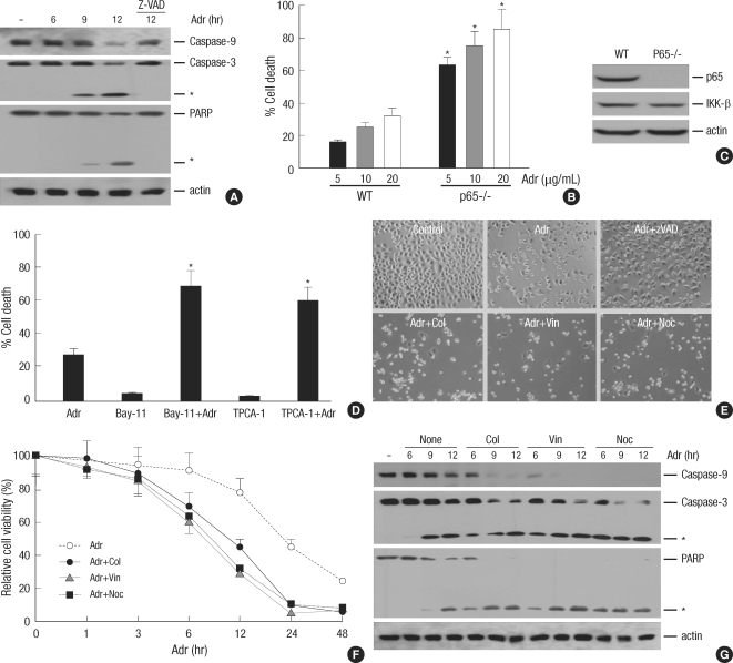
Fig. 5.
Microtubule disrupting agents sensitizes DNA damage-induced apoptotic cell death. (A) HeLa cells were treated with Adr (10 µg/mL) for various times as indicated in the presence or absence of pan-caspase inhibitor z-VAD-FMK (20 µM) and cell extracts were analyzed by immunoblotting with antibodies against casapse-9, -3 and PARP. As a protein loading control, the same amounts of extracts were analyzes by immunoblotting with anti-actin antibody. The asterisks indicate cleaved products of casapse-3 and PARP upon Adr treatment. (B) Wild-type and p65-/- MEF cells were treated with various concentrations of Adr as indicated. 12 hr after treatment, the percentage of cell death was determined by trypan blue exclusion assay as described under Materials and Methods. The results represent the mean values of at least three independent experiments. *, P<0.05, compared with Adr-treated wild-type MEF cells. (C) The expression levels of p65 and IKK-β in wild-type and p65-/- MEF cells. The equal amount of cell extract from each cells was analyzed by immunoblotting with antibodies against p65, IKK-β and actin. (D) HeLa cells were pretreated with the NF-κB specific inhibitors, 5 µM Bay-11 or 1 µM TPCA-1 for 30 min and then treated with 10 µg/mL Adr for 12 hr. Cell death was quantified as described in (B), and each column shows mean±S.E. of at least three independent experiments. *, P<0.05, compared with Adr-treated group. (E) HeLa cells were treated with Adr (10 µg/mL) for 15 hr in the presence or absence of Col (10 µM), Vin (1 µM) and Noc (0.5 µM), or z-VAD-FMK (20 µM). Then, cells were visualized with a normal light microscope with an inverted microscope. (F) HeLa cells were treated with Adr (10 µg/mL) for various times as indicated in the presence or absence of Col (10 µM), Vin (1 µM) and Noc (0.5 µM). Cell death was quantified as described in (B), and each column shows mean±S.E. of at least three independent experiments. (G) HeLa cells were treated as described in (C). Cell extracts form each sample were analyzed by SDS-PAGE followed by immunoblotting with antibodies against casapse-9, -3, PARP and actin. The asterisks indicate cleaved products of casapse-3 and PARP.

