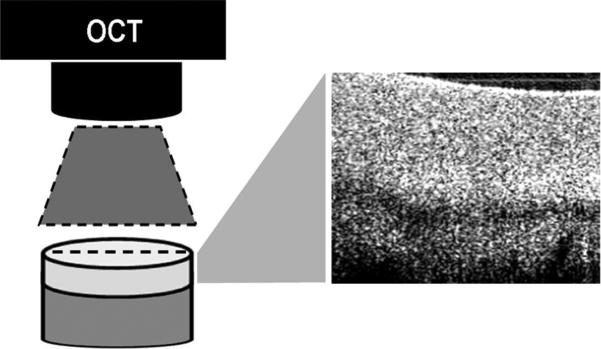
Figure 1.
OCT scanner generates a 2D cross-sectional image (6.5mm x 2mm) along the scan line depicted by the laser aiming light (dashed line). The OCT scan line defines the cartilage section in the mid-sagittal plane running from the 12 o'clock mark to the 6 o'clock position and all cores were scanned in this position in order to preserve the orientation of the acquired OCT images both before and after impact.

