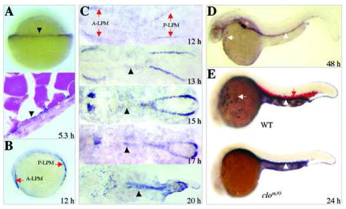
Figure 3.

Spatio-temporal expression of cebpa during zebrafish embryogenesis. (A) At the 50%-epiboly stage (5.3 hpf), expression of cebpa is first detected in the yolk syncytial layer (YSL) at the margin of the blastoderm (top, arrowhead). Cells expressing cebpa are seen in a representative section across the YSL (bottom. arrowhead). (B) At the 6-somite stage (12 hpf), cebpa expression can be seen as distinct stripes, one is located in the anterior lateral plate mesoderm and the other in the posterior lateral plate mesoderm (A-LPM and P-LPM, arrows). (C) Dynamic expression of cebpa in cells of A- and P-LPM during the segmentation period (stages 12 to 20 hpf). (D) At 48 hpf, expression of cebpa can been observed in anterior myeloid cells (arrow), liver (star) and developing gut primordium (arrowhead). (E) At 24 hpf, detection of cebpa (white arrow for myeloid cells; white arrowhead for gut) and α-hemoglobin (red arrow) in wild-type sibling (top) as well as the cloche mutant (clom39, bottom). Note that the cloche mutant lacks both cebpa and α-hemoglobin in hematopoietic cells, but maintains the gut expression of cebpa. Panels A, B, D and E are lateral views, anterior to the left in B, D and E, with animal pole up in A. Panel C shows dorsal views of flat-mounted embryos, anterior to the left.
