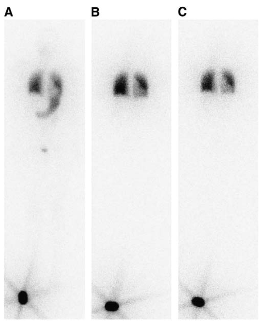
FIGURE 1.
Whole-body planar projection (anterior view) of radioactive iodine distribution 3 h (A), 26 h (B), and 146 h (C) after diagnostic administration of 37 MBq. A standard (~18.5 MBq) was placed by the patient’s right foot during scan. Stomach and bladder can be seen on 3-h scan. Activity is localized to both lungs and retained there 146 h after injection. Gray level intensity in part of left lung is weaker due to overlap of heart.

