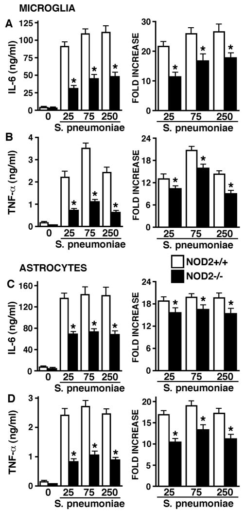
FIGURE 1.
Inflammatory cytokine responses of glia to intact S. pneumoniae are significantly lower in the absence of NOD2 expression. Microglia (Panels A and B) or astrocytes (Panels C and D) (2 × 10⁶ cells per well) from wild type (NOD2+/+) and NOD2 knockout (NOD2-/-) animals were untreated or exposed to viable S. pneumoniae (MOI, of 25:1, 75:1, 250:1 bacteria to each glial cell). At 24 hrs following bacterial challenge culture supernatants were isolated and assayed for the presence of IL-6 (Panels A and C) or TNF-α (Panels B and D) by specific capture ELISA. Data are presented as the culture supernatant cytokine concentrations (Left panels) and as fold increases over levels in unstimulated cells (Right panels) and are the means of triplicate determinations of samples from three separate experiments +/- SEM. Asterisks indicate statistically significant differences in cytokine production between cells derived from wild type and NOD2 deficient animals (p < 0.05).

