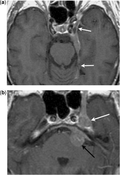
Figure 2.
Centripetal perineural spread. Axial contrast-enhanced T1-weighted images show (a) an en-plaque meningioma along the lateral wall of the left cavernous sinus and tentorial leaf (arrows), with (b) centripetal perineural spread to the trigeminal ganglion in the Meckel cave (white arrow) and the cisternal segment of the trigeminal nerve (black arrow).

