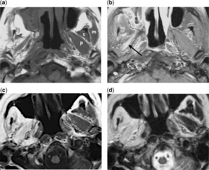
Figure 4.
Denervation atrophy of the masticator muscles. (a) Axial T1-weighted image of a patient with perineural tumour spread along the right mandibular nerve shows significant atrophy of the right masticator muscles, compared with the normal contralateral masseter (m), temporalis (t) and pterygoid (p) muscles. (b) Axial contrast-enhanced fat-suppressed T1-weighted image shows enhancement of the denervated muscle, notably the pterygoid muscles (arrow). (c) Axial T1-weighted and (d) axial T2-weighted images of another patient with similar perineural tumour involvement of the right mandibular nerve show marked fatty infiltration of the right masticator muscles without significant loss of the muscle bulks.

