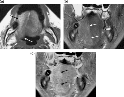
Figure 5.
A 57-year-old male with nasopharyngeal carcinoma spreading along the right hypoglossal nerve resulting in subacute denervation atrophy of the ipsilateral tongue. (a) Axial T1-weighted image shows atrophy of the right side of the tongue with early fatty infiltration (black arrow) and posterior displacement (white arrow). (b) Coronal fat-suppressed T2-weighted image shows hyperintensity in the right side of the tongue due to relative increase in extracellular water (arrows). (c) Coronal contrast-enhanced fat-suppressed T1-weighted image shows enhancement of the right side of the tongue due to underlying increase in perfusion.

