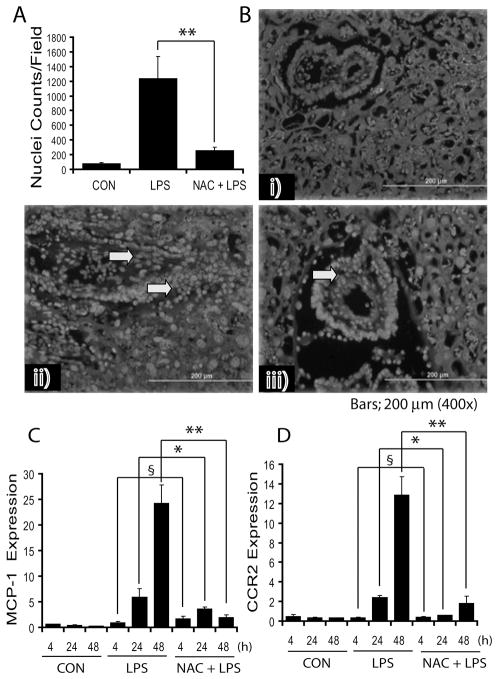
Figure 3. NAC attenuates LPS-induced leukocyte infiltration and chemokine expression in the placenta of pregnant rats.
Plot depicts Hoechst+ nuclei (A), representative image of placental sections show cellular-infiltration [control (i), LPS (ii) and NAC + LPS (iii)] (B). Arrowhead indicates infiltrates in the placental tissue. Plots depict MCP-1 (C) and CCR2 (D) transcripts in the placentas of pregnant rats (n = 6) following LPS exposure in the presence/absence of NAC. Results in plots are expressed as Mean ± SD. Statistical significance * p<0.05, ** p<0.001 and non-significant (§). Magnifications at 400 ×s.

