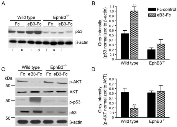
Figure 6.
EphB3 directly regulates p53 and AKT phosphorylation in the SVZ. EphB3 was stimulated in vivo by infusing pre-clustered ephirnB3-Fc (eB3-Fc) or Fc-control (Fc) molecules into the lateral ventricle of wild type and EphB3−/− mice. (A) Western blot analysis of p53 protein expression 3 days after eB3-Fc and Fc infusions show an increase in p53 expression in the ipsilateral (i) and contralateral (c) SVZ of wild type but not EphB3−/− mice. (B) Bar graph representing quantified data of p53 normalized to β-actin control levels in the SVZ of wild type and EphB3−/− mice (n=4). (C) The level of phosphorylated AKT (p-AKT) is decreased in the ipsilateral SVZ 3 days after eB3-Fc stimulation as total p53 and phosphoSer15-p53 are increased in wild type while having no effect in EphB3−/− mice. (D) Bar graph representing quantified data of p-AKT levels normalized to total AKT levels in the SVZ of wild type and EphB3−/− mice (n=4). *P<0.05 and **P<0.01 compared to wild type Fc-control.

