
FIGURE 2.
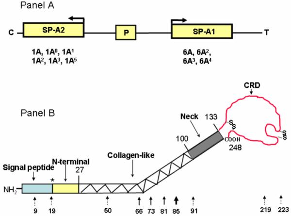
Schematic presentation of the human SP-A locus and the protein domains and genetic variations of SP-A. Panel A depicts the organization of the human SP-A locus (SP-A2, pseudogene [P], and SP-A1), the transcriptional orientation (arrow) of each gene, and the corresponding, more frequently observed coding variants below each gene. Panel B depicts the various protein domains of SP-A and the position of each of the 10 collective amino acid differences among the human SP-A variants (reviewed in Ref. 20). The number above the structure shows the numbers of the last amino acid of the SP-A precursor where the preceding domain ends. * denotes that the signal peptide could be 18, 19, or 20 amino acids. The number below the structure indicates the amino acid number of the SP-A precursor. Solid-line arrows denote the gene-specific differences that distinguish all SP-A1 variants from the SP-A2 variants. The dotted-line arrows show differences among variants of either gene.