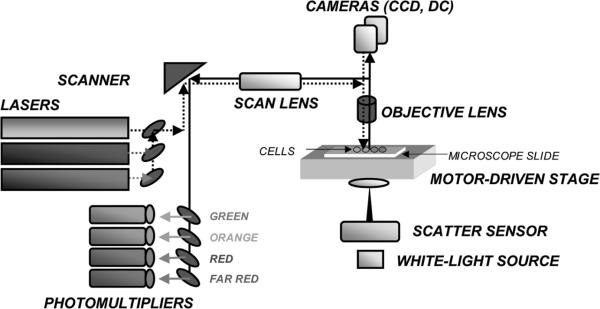
Figure 4.
Schematic representation of the laser scanning cytometer (LSC). The microscope is the key part of LSC and it provides structural and optical components. The emission beams from lasers are directed onto computer controlled oscillating mirror, which reflects them through the epi-illumination port of the microscope and images through the objective lens onto the slide. The mirror oscillations cause the laser beam to sweep the area of microscope slide under the lens. The slide is located on the computer-controlled motorized microscope stage which moves perpendicular to the laser beam scan at 0.5 μm steps per each scan. The cell-emitted fluorescence is collected by the objective lens and directed to the scanning mirror. Upon reflection it passes through a series of dichroic mirrors and emission filters to reach one of the PMTs, which records the fluorescence intensity at a specific wavelength range. Laser light scattered by the cell is imaged by the condenser lens and its intensity is recorded by sensors. A white-light source provides transmitted illumination to visualize the objects though an eyepiece or cameras. Up to 100 cells can be analyzed per second with accuracy and sensitivity comparable to that of flow cytometry (Fig. 3). A color version of the figure is available at www.landesbioscience.com/curie.

