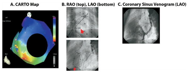
Figure 3.
A. Three dimensional electroanatomical map (CARTO) of retrograde atrial activation during SVT as seen in the RAO projection. The area of earliest activation (in red) occurs on the anatomic tricuspid valve annulus in the posterolateral region. B. Site of successful ablation. RAO and LAO projections show the distal tip of the ablation catheter (marked by arrow) between the 7–8 o’clock position on the tricuspid valve annulus. Other reference catheters include a coronary sinus catheter, a His catheter, and a RV catheter. C. Coronary sinus (CS) venogram in LAO projection, after injection at the mouth of the CS; it shows opacification of the mouth of CS around the posterior mitral valve and the lesser cardiac vein running around the posterior tricuspid valve.

