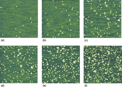
Figure 11.
Series of consecutive AFM images (1 μm×1 μm) of BSA (20 μl of 0.1 mM protein solution in acetate buffer of pH 4.75 and ionic strength I=8 mM at room temperature) adsorbed onto mica taken at the same area (slight drift) under stopped flow conditions. The bright objects represent single proteins.

