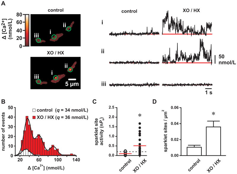
Figure 2. Reactive oxygen species increase L-type Ca2+ channel sparklet activity in isolated cerebral arterial smooth muscle cells.
A, Representative TIRF images showing Ca2+ influx in an arterial myocyte before and after application of XO and HX (2mU/mL and 250 μmol/L, respectively). Traces show the time course of Ca2+ influx at the three circled sites before and after XO/HX. B, Ca2+ sparklet amplitude histograms before (white) and after XO/HX exposure (red). The solid black lines are best-fits to the control (q=34 nmol/L) and to the XO/HX (q=36 nmol/L) histograms with a multi-component Gaussian function where q is the quantal unit of Ca2+ influx (see Detailed Methods in the Online Supplement). C, Plot of Ca2+ sparklet site activities (nPs) before and after XO/HX (n=8 cells). The red solid lines are the arithmetic means of each group and the dashed line marks the threshold for high-activity Ca2+ sparklet sites (nPs≥0.2). D, Plot of the mean ± SEM Ca2+ sparklet density (Ca2+ sparklet sites/μm2) before and after XO/HX (n=8 cells). *P<0.05

