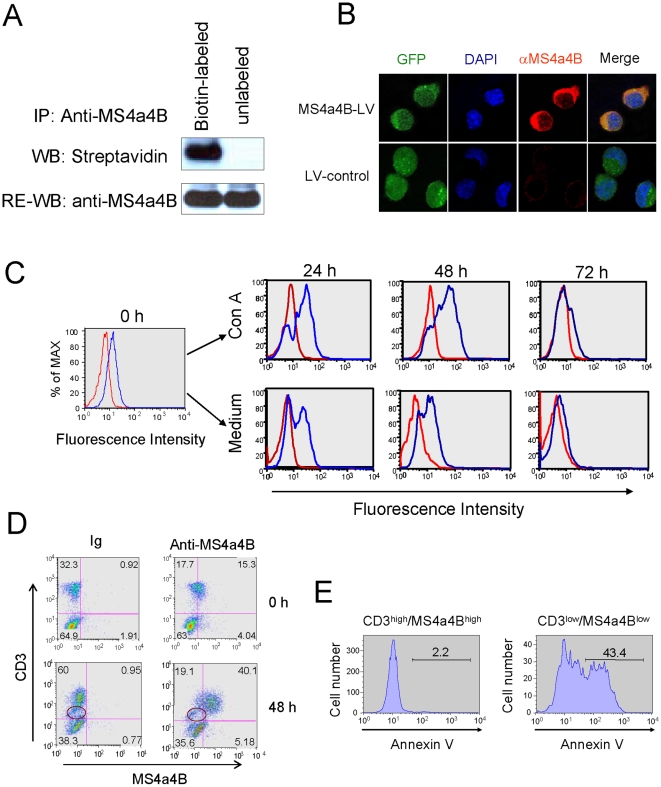
Figure 1. MS4a4B is expressed on cell surface and is potentially associated with activation of T cells.
A, To determine whether MS4a4B is expressed on cell surface, mouse spleen cells were surface-labeled with EZ-Link Sulfo-NHS-SS-Biotin according to the manufacturer's protocol (Pierce). Unlabeled spleen cells were used as control. Cells were lysed by lysis buffer containing 1% NP-40. Cell lysate was immunoprecipitated by anti-MS4a4B-coupled protein A-beads and was separated on 12% SDS-PAGE, followed by blotting with streptavidin-HRP. MS4a4B was confirmed by re-blotting with anti-MS4a4B antibody. B, EL4 cells, infected with either MS4a4B-expressing lentivirus (LV) vector or mock LV vector, were stained with rabbit anti-MS4a4B antibody followed by labeling with anti-rabbit-IgG-Cy3 conjugate. Expression and localization of MS4a4B were observed by confocal microscopy. Magnification, ×40. C, Spleen cells were cultured for 24, 48 and 72 hrs in the presence or absence of Con A (5 µg/ml). Spleen cells before culture were used as control (0 hr). Cells were first stained with anti-CD3-PE, and then intracellular stained with biotinylated anti-MS4a4B antibody (blue line) or biotinylated Ig control (red line), followed by labeling with streptavidin-Red 670. For flow cytometric analysis, cells were first gated on CD3, and then were analyzed for MS4a4B expression. D, Spleen cells pre- or 48 hr post Con A stimulation were co-immunostained with anti-CD3 and anti-MS4a4B antibody. Cells were analyzed by flow cytometry. Note the CD3low/MS4a4Blow population. E, Spleen cells were stimulated with Con A for 48 hr and were co-stained with Annexin V, anti-CD3 and anti-MS4a4B antibodies. CD3low/MS4a4Blow and CD3high/MS4a4Bhigh populations were gated for assessment of apoptosis indicated by Annexin V binding.

