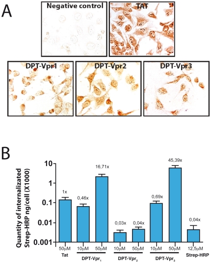
Figure 2. Effect of DPT-Vpr peptides on cell penetration and intracellular delivery of streptavidin-peroxydase.
(A) For penetration and localization analysis, cells were incubated with 150 µM of peptides for 2 h at 37°C. After fixation, the presence of biotinynlated peptides is revealed by incubation of permeabilized cells with streptavidin-peroxydase. The sequence of non penetrating peptide used as negative control is GVIFYLRDK. The sequence of positive Tat control is YGRKKRRQRR. (B) Intracellular delivery of streptavidin-peroxydase by biotinylated-DPT-Vpr and Tat peptides in HeLa cells. Streptavidin-peroxydase coupled with biotinylated peptides were incubated for 6 h at 37°C and internalized complexes were visualized by a colorimetric test. Statistical analysis was carried out using Anova's test and significance was assessed at p<0.0001.

