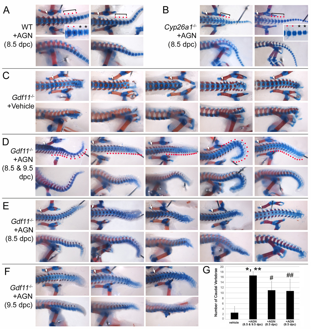
FIG. 7.
Tail truncation defects of Gdf11−/− mice are significantly rescued by a pan retinoic acid receptor antagonist, AGN193109 (AGN). Representative ventral (upper panels) and lateral (lower panels) views of posterior lumbar, sacral, and proximal caudal vertebrae are shown. All skeletons were collected from E18.5 fetuses, except for Cyp26a1−/− from E17.5 (B). White arrows indicate the first sacral vertebra. (A) WT treated with AGN. Red dots with a bracket indicate proximal caudal vertebrae having transverse and spinous processes. Asterisks indicate caudal vertebrae that do not contain transverse and spinous processes. (B) AGN-treated Cyp26a1−/− skeletons. AGN treatment almost completely rescued the caudal agenesis defect of Cyp26a1−/− fetuses. The morphological transition in the caudal vertebrae is also restored (inset). (C) Vehicle-treated Gdf11−/− skeletons. (D) Gdf11−/− skeletons treated with AGN for two consecutive days at 8.5 and 9.5 dpc. AGN-treated Gdf11−/− embryos show elongated tail. Take notice that the extended caudal vertebrae contain transverse and spinous processes (indicated by red dots). (E, F) Gdf11−/− skeletons treated with AGN at 8.5 dpc (E) and 9.5 dpc (F). (G) Histogram showing the number of caudal vertebrae, defined as vertebral segments present posterior to 4th sacral vertebra. Means and standard deviations are shown as filled box and bar above each bar. * (p < 0.0001), # (p < 0.001), and ## (p < 0.01): compared with vehicle-treated Gdf11−/− embryos as determined by Student's t test. ** (p < 0.05) compared with AGN-treated Gdf11−/− embryos at 8.5 or 9.5 dpc.

