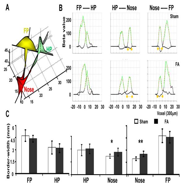
Figure 5. Analysis of the 1D spatial profile of BOLD response along lines across CMs.
Three lines cross CMs of forepaw, hindpaw, and nose are demonstrated in the schematic graph (A). The beta value of each voxel along the line was plotted as a function of space in the 1D profile (B). Two lines were projected from each CM, and a total of six 1D spatial profiles were analyzed (A, FP, yellow; HP, green; nose, red). The filtered 1D spatial profile (green) was fitted by a second-order polynomial curve (red) for each line (B). The border-width from the extrapolated borders (B, orange dots) along each line was calculated to estimate the spatial extent of the BOLD response. A significantly expanded BOLD response in the S1 representation of nose in F.A. rats is shown (C). (* means p = 0.01;** p = 0.002; two tail, NSHAM = 6, NF.A.= 9).

