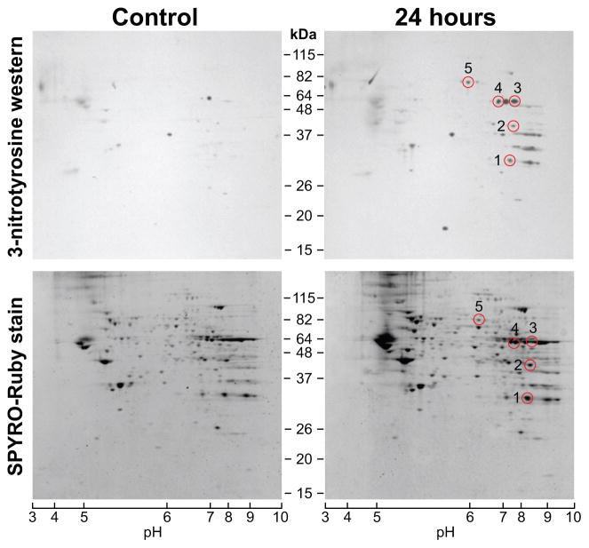
Figure 3.
Two-dimensional analyses of mitochondrial proteins from hippocampi of rats after acute exposure to combustion smoke. 2DE profiles of mitochondrial proteins and corresponding detection of 3-nitrotyrosine (3-NT) immunoreactive mitochondrial proteins. Mitochondrial proteins from controls and rats at 24 h after smoke were resolved in the first dimension by IEF (pH range 3–10) followed by separation in a gradient 8–16% SDS-PAGE. The bottom panels show protein profiles visualized by SYPRO-Ruby protein stain for sham-controls and rats 24 h post smoke. Corresponding Western blots of parallel gels electrotransferred to PVDF membranes and probed for 3-NT are given in top panels. Western blotting reveals a distinct pattern of nitrated proteins when compared to controls. SYPRO-Ruby spots were aligned with Western blot signals (circles), excised, in gel digested with trypsin and analyzed by MS. Nitrated protein spots identified with high confidence are listed and described in Table 1.

