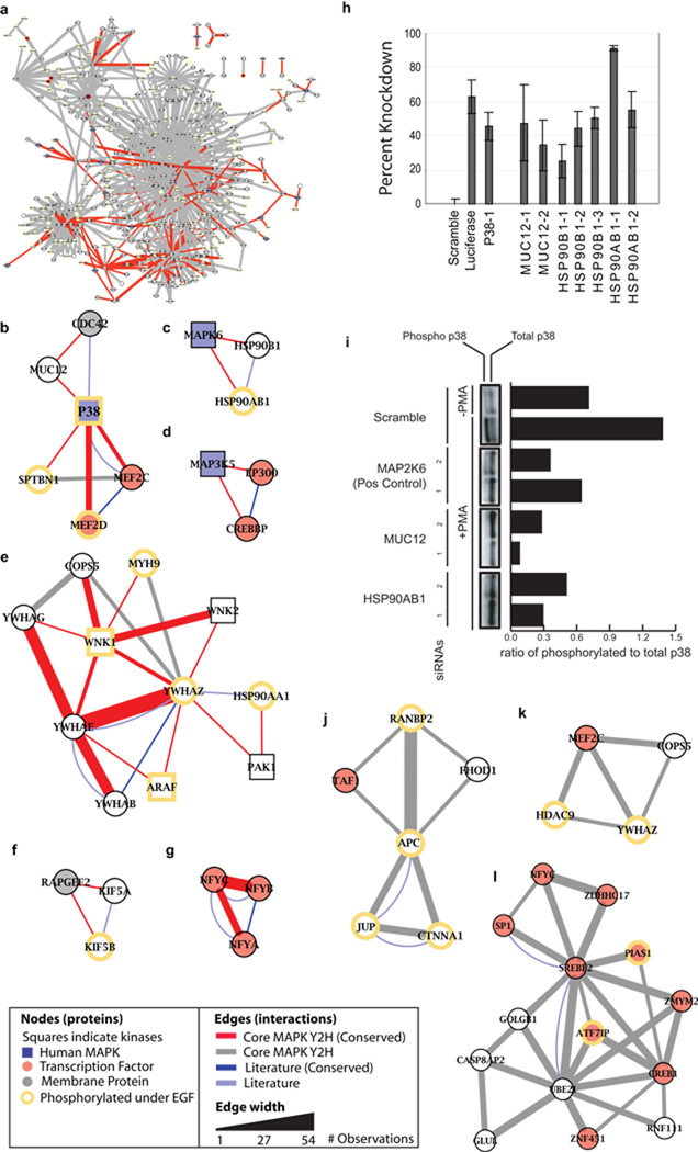
Figure 3.
Functional modules in the core network. (a) Bird’s eye view of the core MAPK Y2H network. (b–g) High confidence conserved functional modules. Red edges correspond to core MAPK Y2H interactions which were conserved with yeast. Grey edges indicate core interactions not conserved with yeast. Thickness of the edge increases with the number of observations. (h) AP-1 luciferase activation assay for various siRNAs targeting members of conserved modules. (i) p38 phosphorylation levels are decreased with siRNAs targeting members of conserved modules. (j–l) Novel modules not conserved with yeast.

