
Figure 4.
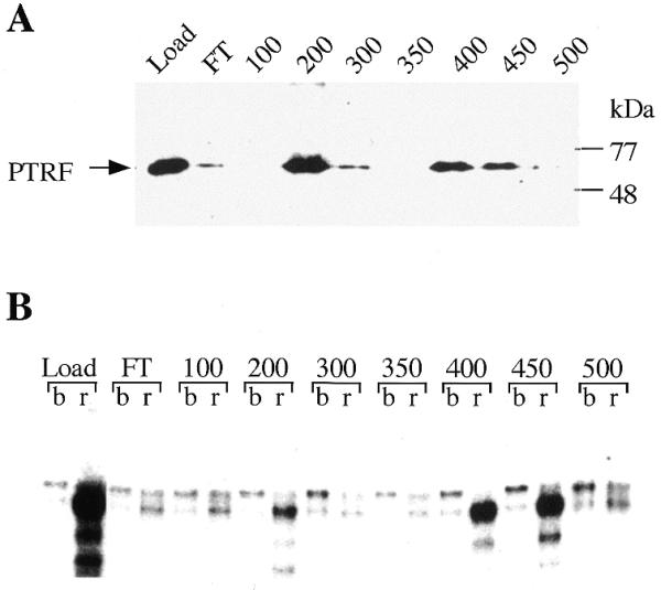
Chromatographic separation of active and inactive forms of PTRF. (A) Western blot. PTRF-containing fractions from a phosphocellulose column were pooled (Load) and fractionated on S-Sepharose. The flow-through (FT) and individual fractions eluting at the salt concentrations indicated were analyzed on western blots using chicken anti-PTRF antibodies. (B) Transcript release assay. The reactions contained bead-bound 3′-end tailed pCAT-T6-T1, 0.5 U Pol I, 30 ng TTF-I and 5 µl of the respective fractions shown at the top of the gel. After incubation, the assays were separated into template-bound (b) and released (r) fractions.