
Fig. 2.
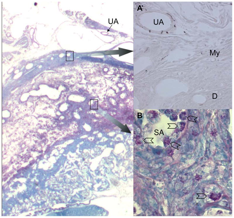
Photomicrographs of serial cryostat sections of gd10 C57Bl/6J implantation sites. Left panel shows Periodic Acid Schiff’s stained section with methylene blue counterstain, imaged at 40x. Insert (A) shows enlargement (imaged at 200x) of the upper boxed region with immunolocalization of sympathetic nerves using a primary goat antibody to tyrsoine hydroxylase (Chemicon, Th42), a rabbit anti-goat secondary reagent and DAB as the chromogen. Strong staining of sympathetic nerve fibres is seen in the walls of branches from the uterine artery (UA) as they pass in the mesometrium towards the uterus. Immunoreactivity is lost in the myometrium (My) and is absent from the decidua (D) and the walls of the spiral arterioles (SA) in the decidua x 400. Insert (B) shows enlargement (imaged at 400x) of the lower boxed region. UNK cells (chevrons) are abundant in the decidua and surrounding and within the walls and lumens of the spiral arteries. No evidence for co-localization of nerves and uNK cells was seen between gd6-12.