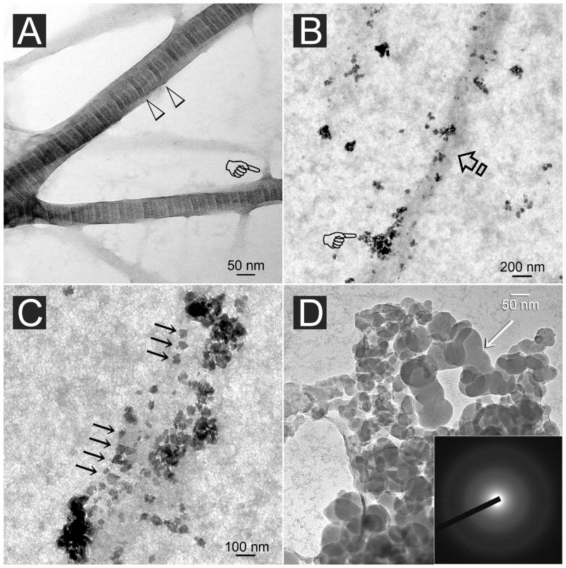
Fig. 5.
A. Stained image of reconstituted type I collagen showing an approximately 76 nm periodicity (open arrowheads) in fibrils that are larger than 50 nm in diameter. Branching of the fibrils could frequently be observed (pointer). B. Unstained, PVPA-immobilized collagen fibril (arrow) retrieved after 4 h of immersion in the mineralization medium (see text). Electron-dense nanospheres (pointer) aggregated along the periphery of the unmineralized fibril. C. Unstained image showing attachment of electron-dense nanospheres along an unmineralized fibril with a periodicity (arrows) that roughly corresponded with the D-period of a stained fibril. D. High magnification of the partially coalesced nanospheres (arrow) illustrating the fluidity of the nanoprecursor phase. Inset: Electron diffraction revealed a diffuse pattern characteristic of the amorphous nature of the initially formed minerals.

