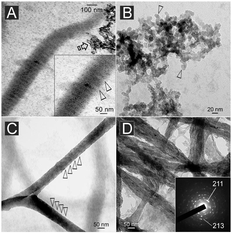
Fig. 7.
A. Unstained reconstituted collagen fibrils cross-linked with EDC to immobilize 500–1,000 μg/mL of PVPA within the fibrils and retrieved after incubation in the mineralization medium for 24 h. Electron-dense intrafibrillar minerals were deposited in a manner that revealed the periodicity of the fibrils (inset: between open arrowheads). B. Unstained image of the aggregates depicted by the open arrow in A revealed formation of angular nanocrystals with roughly hexagonal appearance (open arrowheads). C. Unstained, highly mineralized reconstituted collagen fibrils that were retrieved after 24 h. Elevations along the periphery of the mineralized fibril revealed a vague periodicity (open arrowheads). D. Unstained image of similar mineral fibrils showing the formation of electron-dense nanocrystals within the fibrils. Internal periodicity could not be identified due to the three-dimensional nature of the fibrils. Inset: concentric ring patterns with arc-shaped patterns in the (211) plane that are indicative of the formation of apatite with a regular orientation (i.e. parallel to the longitudinal axis of the collagen fibril).

