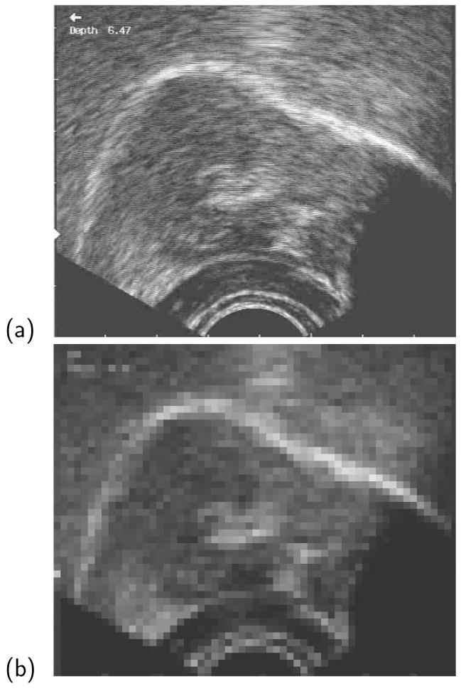
Figure 4.

Example frame from recorded ultrasound, showing the region representing tongue activity (a) as recorded and (b) pixelized prior to the calculation of Delta. The tongue root is on the left and the tongue-tip on the right of the image. Note that Depth information and other indicators in the image are invariant, and the corresponding pixels therefore contribute zero to calculations of Delta.
