Fig. 1.
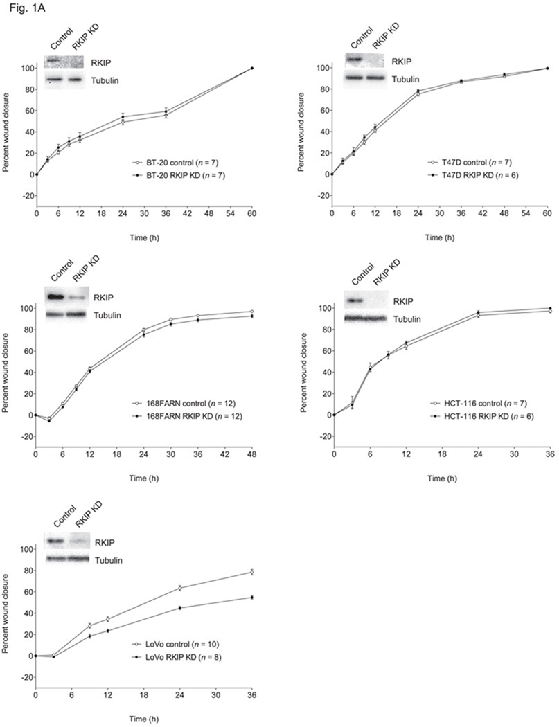
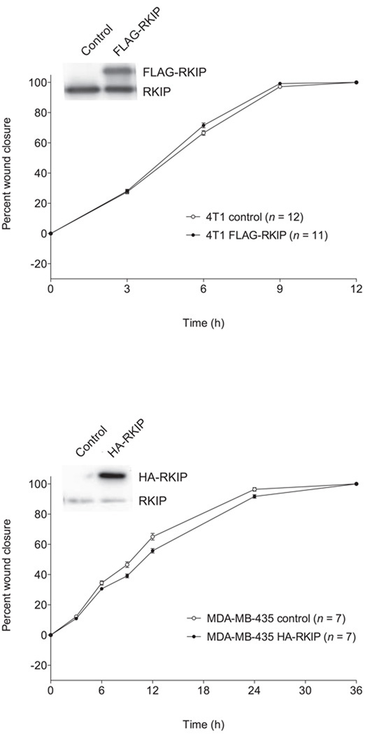
Effects of altered RKIP expression on migration of different cancer cell lines. The values represent the means and standard errors of the mean (SEM) for the percent closure of wounds in cell monolayers as a function of time (n = indicated number of separately treated wounds on multiwell tissue culture-treated plates from three independent experiments). (A) BT-20, T47D and 168FARN breast cancer cells and HCT-116 and LoVo colon cancer cells stably expressing a control siRNA for firefly luciferase (control) and those expressing an RKIP-specific siRNA (RKIP KD). Above the graphs are representative Western blots showing expression of RKIP and α-tubulin in the corresponding cell lines. (B) Control and RKIP-overexpressing 4T1 breast cancer cells and MDA-MB-435 melanoma cells (stably expressing FLAG-RKIP and HA-RKIP, respectively). Above the graphs are representative Western blots showing expression of tagged and endogenous RKIP in the corresponding cell lines.


