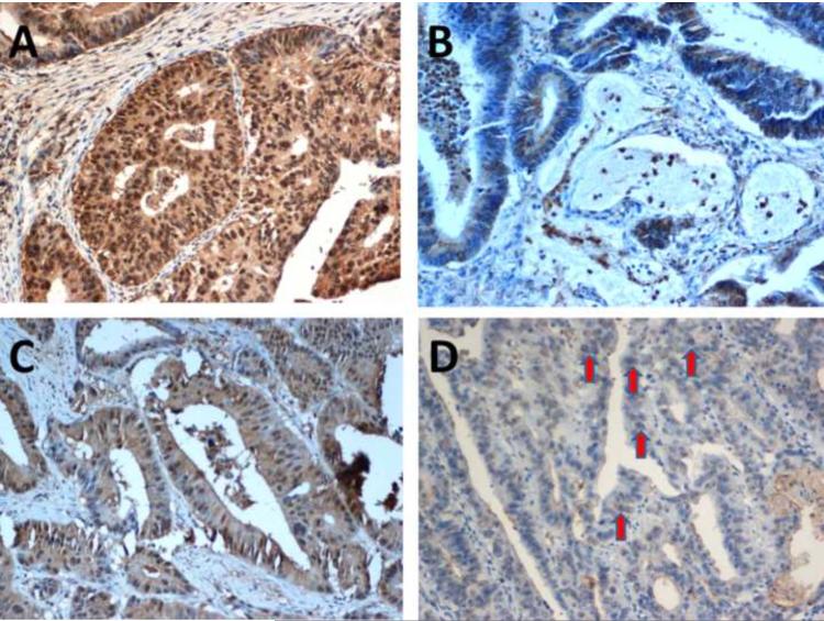
Figure 2. hMSH3 expression patterns by immunohistochemistry.
(A) An hMSH3-positive (brown nucleus staining) tumor. (B) An hMSH3-negative (blue nucleus staining) tumor. Most EMAST tumors showed (C) a mixed appearance with partial hMSH3 loss. (D) Heterogeneity was diagnosed when there is a nucleus showing both brown and blue staining in the nucleus (arrows).

