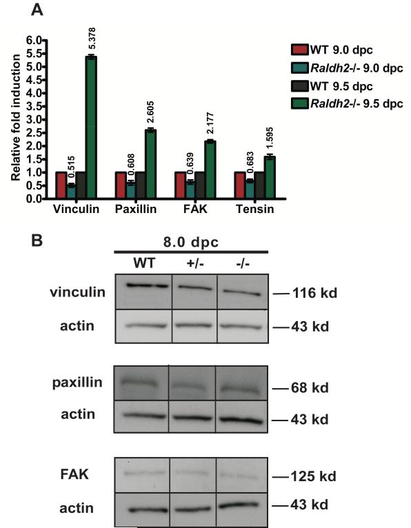
Figure 4. Regulation of focal adhesion formation and turnover is critical during the vascular remodeling stage of blood vessel development (9.5 dpc).
(A) mRNA expression of vinculin, paxillin, FAK, and Tensin1 was assessed by qRT-PCR in yolk sacs harvested at 9.0 dpc (15-20 somite stage) versus 9.5 dpc (25-30 somite stage). Relative to corresponding WT yolk sacs isolated from littermates at either 9.0 dpc or 9.5 dpc, mRNA expression of these genes is increased in Raldh2−/− yolk sacs specifically at 9.5 dpc and not prior at 9.0 dpc. Transcript levels were normalized by using β-actin as the internal control. (B) Protein levels of FAK, paxillin, and vinculin were determined by Western blot in 8.0 dpc WT, Raldh2 heterozygote (+/−), and Raldh2 null (−/−) yolk sacs isolated at the 3-6 somite stage prior to cardiac function and establishment of blood flow. At this stage, protein levels in Raldh2 −/− yolk sacs were not found to be different from WT or Raldh2 +/− yolk sacs. Actin was used as the loading control.

