Abstract
Exposure of pheochromocytoma (PC12) cells to hypoxia (1% O2) favors differentiation at the expense of cell viability. Additional incubation with nerve growth factor (NGF) and guanosine, a purine nucleoside with neurotrophin characteristics, rescued cell viability and further enhanced the extension of neurites. In parallel, an increase in the activity of protein kinase C-related kinase (PRK1), which is known to be involved in regulation of the actin cytoskeleton, was observed in hypoxic cells. NGF and guanosine further enhanced PRK1 in normoxic and hypoxic cells. To study the role of PRK1 during cellular stress response and neurotrophin-mediated signaling, PC12 cells were transfected with small interfering RNA (siRNA) directed against PRK1. Loss of functional PRK1 initiated a significant loss of viability and inhibited neurite formation. SiRNA-mediated knockdown of PRK1 also completely stalled guanosine-mediated neuroprotective effects. Additionally, the F-actin-associated cytoskeleton and the expression of the plasticity protein GAP-43 were disturbed upon PRK1 knockdown. A comparable dependency of neurite formation and GAP-43 immunoreactivity on functional PRK1 expression was observed in cerebellar granule neurons. Based on these data, a putative role of PRK1 as a key-signaling element for the successive NGF- and purine nucleoside-mediated protection of hypoxic neuronal cells is hypothesized.
Keywords: Protein kinase C-related kinase 1, hypoxia, neuroprotection, guanosine, PC12 cells, cerebellar granule neurons
Multiple signaling pathways regulate the critical balance between cell death and survival in ischemia–reperfusion. Even brief episodes of hypoxia can result in cessation of oxidative phosphorylation and depletion of cellular ATP, which results in profound deficiencies in cellular function. As a consequence, sustained hypoxia can lead to cell death. Following ischemia, dying, injured, and hypoxic cells release soluble nucleotide and nucleoside pools (Neary et al. 1996), which are then further metabolized to other purine derivatives (Zetterstrom et al. 1982) that remain elevated for days after the insult (Uemura et al. 1991). Growing evidence suggests that nucleotides and nucleosides might also act as trophic factors in both the central and peripheral nervous systems. Specific extracellular receptor subtypes for these compounds are expressed on neurons, glia, and endothelial cells, where they mediate strikingly different effects. Such effects range from induction of cell differentiation, apoptosis, mitogenesis, and morphogenetic changes, to stimulation of synthesis and/or release of cytokines and neurotrophic factors under both physiological and pathological conditions. Nucleotides and nucleosides are therefore likely to be involved in the regulation of the nervous system’s development and plasticity (Neary et al. 1996).
We have previously studied neuronal signaling after chemical hypoxia and observed the protective capacity of purine nucleosides in PC12 cells (Tomaselli et al. 2005a; Tomaselli et al. 2005b) and in primary cerebellar granule neurons (Bocklinger et al. 2004; Heftberger et al. 2005). Among purine nucleosides, guanosine gained our special attention due to its neurite stimulating capacity. Guanosine, a purine nucleoside present in the brain under both physiological and pathological conditions (Zetterstrom et al. 1982; Uemura et al. 1991), has many trophic and neuroprotective effects (Rathbone et al. 1998; Ciccarelli et al. 2000; for review see Schmidt et al. 2007), and guanine-based purines synergistically enhance NGF-dependent neurite outgrowth (Gysbers and Rathbone 1996; Tomaselli et al. 2005b). Earlier data indicated a role for protein kinase C-related kinase 1 (PRK1, also known as PKN or PKNα) in cellular stress responses (Tomaselli et al. 2005a). PRK1 is a lipid-activated serine/threonine protein kinase and a member of the protein kinase C (PKC) superfamily (Mellor and Parker 1998; Mukai 2003) of potential key regulators orchestrating physiological responses, and is involved in regulation of the actin cytoskeleton. PRK1 is activated by interacting with the Rho and Rac families of small G proteins and arachidonic acid, or by caspase cleavage (Vincent and Settleman 1997; Takahashi et al. 1998; Lu and Settleman 1999; Mukai 2003), and has been shown to interact with alpha-actinin and to promote neuronal differentiation (Gudi et al. 2002).
Published literature and our own data stimulated our further interest in the study of a potential role for PRK1 and guanosine in the endogenous mechanisms involved in the protection of neuronal cells that are subjected to hypoxic insult. We also sought to clarify specific intracellular adaptations that contribute to changes in the plasticity of these cells following trauma and ischemia (Tomaselli et al. 2005a). O2-sensitive (Zhu et al. 1996; Seta et al. 2002) clonal rat PC12 cells, a widely utilized model for sympathetic ganglion-like neurons (Greene and Tischler 1976) were used. PC12 cells express abundant A2A adenosine receptors (A2AR) (Hide et al. 1992; van der Ploeg et al. 1996; Arslan et al. 1999; Tomaselli et al. 2005b), which effect these cellular responses to hypoxia (Kobayashi et al. 1998; Kobayashi and Millhorn 1999). Data were verified in postnatal day 7 cerebellar granule neurons, a primary rat cell model that appeared suitable for morphological and biochemical studies following exposure to low oxygen and were previously established for the study of purinergic signaling (Bocklinger et al. 2004; Tomaselli et al. 2008; Zur Nedden et al. 2008).
Materials and methods
Materials
PC12 cells were ordered from LGC Promochem ATCC (Manassas VA, USA). RPMI 1640 medium, DMEM, L-glutamine, and penicillin/streptomycin were purchased from PAA Laboratories (Vienna, Austria). Neurobasal medium, the B27 supplement, trypsin, trypsin inhibitor, horse serum, and fetal calf serum were from GIBCO Invitrogen (Vienna, Austria). NGF-ß, purine nucleosides (guanosine), poly L-ornithine, bovine serum albumin (BSA), aprotinin, leupeptin, NaF, NaP-P, Na3VO4, paraformaldehyde (PFA), propidium iodide (PI), Phalloidin-FITC, and anti-pan-GAP43 clone GAP-7B10 were obtained from Sigma (Cologne, Germany). All culture flasks, dishes, and collagen-S type I were from Becton Dickinson (Canaan CT, USA). chamber slides were from NUNC (Rochester NY, USA) and glass cover slips were from Assistant (Sondheim, Germany). DNase I was purchased from Roche (Mannheim, Germany). Hoechst 33342 and TRITC- or FITC-conjugated goat anti-mouse/rabbit IgG F(ab’)2 fragments for immunostaining were obtained from Molecular Probes (Oregon, USA). Percoll, nitrocellulose membrane, and Hyper-ECL film were obtained from GE Healthcare Biosciences (Uppsala, Sweden). Mounting medium for immunofluorescence studies was from Dako (Vienna, Austria). The siGENOME SMART pools for PRK1 and the pool of scrambled non-targeting small interfering RNA (siRNA) duplexes were purchased from Dharmacon (Chicago IL, USA). The enhanced chemiluminescence HRP-substrate (ECL-reagent) was obtained from Millipore (Vienna, Austria). Anti-phospho-PRK1, anti-pan-PRK2, and anti-pan-AKT were obtained from Cell Signaling Technology (Danvers MA, USA). Anti-pan-PRK1 was obtained from BD Biosciences (San Jose CA, USA). Goat anti-rabbit IgG or goat anti-mouse IgG HRP-linked antibodies were from Pierce (Rockford IL, USA). Further reagents for transfection were obtained from Amaxa Biosystems (Lonza, Cologne, Germany). All other chemicals were from NeoLab (Migge, Germany) or Applichem (Darmstadt, Germany).
Cell culture
Experiments were performed in two different cell models that were optimized in our laboratory (PC12 cells, and primary postnatal day 7 cerebellar granule neurons) although these cell models differ with regard to differentiation stage (Kuhar et al. 1993; Hatten et al. 1997) (e.g., p7 cerebellar granule cells were already more differentiated than our PC12 cells).
PC12 cells were grown in RPMI 1640 medium supplemented with 1% Pen/Strep, 1% L-glutamine, 10% horse serum, and 5% fetal calf serum (described as full RPMI 1640 medium throughout the paper) at 37°C with 5% CO2 on collagen-S type I-coated culture dishes. Cells were subcultured at a density of about 80% for 2 days before onset of an experiment.
Primary cerebellar granule neurons were prepared from the cerebellum of postnatal day 7 (p7) Sprague-Dawley rats according to a published protocol (Hatten 1985), which was slightly modified by our group (Bocklinger et al. 2004; Tomaselli et al. 2008; Zur Nedden et al. 2008). Briefly, the dissected cerebellum was treated with 1% trypsin (15 min, 37°C) and 10mg/ml DNase I for 10 min at room temperature (RT). Isolated cells were further purified by a 35/65% Percoll gradient centrifugation (1258g, no brake, 15 min, 4°C). Cells collected from the interphase were further enriched by a preplating step and finally seeded on poly-L-ornithine-coated dishes in Neurobasal medium enriched with 2% B27 supplement, 1% L-glutamine, and 1% Pen/Strep (described as full Neurobasal medium throughout the paper). This preparation allows a highly pure culture of cerebellar granule cells containing 95% small interneurons (Thangnipon et al. 1983; Hatten 1985), and therefore cells were called granule neurons throughout the paper. P7 cerebellar granule neurons were stimulated after two days in vitro.
Induction of hypoxia in PC12 cells and cerebellar granule neurons
Hypoxia was induced by reduction of the oxygen concentration from 21% to 1%. A special cell incubator designed to maintain a low oxygen level (HERAcell 240, Thermo electron cooperation, Austria) was set to a constant condition of 1% O2, balanced with N2, controlled by O2- and CO2-sensors. Small culture volumes guaranteed fast equilibration of the medium.
Post-transcriptional gene silencing
Synthetic siRNA directed against PRK1 and scrambled non-targeting siRNA duplexes (control siRNA) were used for transient Amaxa Nucleofector-based transfection as described previously (Tomaselli et al. 2008; Zur Nedden et al. 2008). Briefly, 1.3×10⁷ PC12 cells were mixed with nucleofector solution from the “Neuronal Cell NucleofectorTM kit V” and with 100-1000 nM siRNA. After transfection with the nucleofection device I (program U-29), cells were transferred to prewarmed culture dishes. The transfection efficiency of PC12 cells was 79.366% ± 7.833%. For transfection of cerebellar granule neurons, a special protocol for the nucleofection of primary neurons (Gartner et al. 2006) was applied. Freshly isolated neurons (1×10⁷ cells) were transfected with 1 μM siRNA using the “Rat neuron Nucleofector kit” and the program O-03 of the nucleofection device I. Transfected neurons were transferred to DMEM supplemented with 1% L-glutamine, 1% Pen/Strep, and 10% horse serum, and after 3h the medium was replaced by full Neurobasal medium. Twenty-four hours later, the transfection medium was changed and cells were incubated for 1-2 days prior to experiments. Transfection efficiency of cerebellar granule neurons was 72.675% ± 3.580%.
Cell viability assay
Experiments were performed as previously described by our group (Bocklinger et al. 2004; Tomaselli et al. 2008; Zur Nedden et al. 2008). Briefly, wild type or transfected cells were incubated under normoxic (N-co) and hypoxic conditions (H-co). Hypoxic cells were further stimulated once with 500μM guanosine (Guo), 5ng/mL NGF-ß (NGF), or a combination of guanosine and NGF (Guo+NGF). After 6-8h, cell viability was measured by staining the cells with the fluorescent dyes Hoechst 33342 (10μg/mL, 10 min, 37°C), which penetrates both living and dead cells, and PI (5μg/mL, 5 min, 37°C), which is membrane impermeable and stains only the DNA of cells with disrupted plasma membranes. Cells of 3-5 independent microscopic fields under blind trial conditions were visualized on a fluorescence microscope (Zeiss Axioplan2, Austria) equipped with a spot camera (RT-slider 2.3.1 Visitron Systems, Germany). Both fluorescence excitations were merged using Adobe Photoshop7.0 software; dead cells are double stained (pink) and viable ones are blue. Hence, the percentage of cell death [pink cells × 100/total cells] was calculated.
Neurite outgrowth assay
Wild type or transfected cells were incubated under normoxic (N-co) and hypoxic conditions (H-co). 500μM guanosine (Guo), 5ng/mL NGF-ß (NGF), or both (Guo+NGF) were added once to hypoxic cells. After various time periods (wild type cells: 24h; transfected cells: 3 days), pictures were taken from 3-5 independent microscopic fields under blind trial conditions, and neurite-bearing cells were counted. Cells with neurites longer than two times their cell diameter were defined as cells with neurite outgrowth. Percentages of neurite formation were calculated [neurite-bearing cells × 100/total cells].
Analysis of cell morphology (Phalloidin staining)
PC12 cells (wild type or transfected with siRNA constructs), were cultured on chamber slides and treated once with 500μM guanosine (Guo), 5ng/mL NGF-ß (NGF), or both (Guo+NGF) under normoxic (N-co) or hypoxic (H-co) conditions. For cytoskeleton studies, cells were generally stained with phalloidin after 24h treatment unless described otherwise. Briefly, cells were washed carefully with 1x phosphate buffered saline (PBS) and fixed for 10 min at RT in 4% PFA supplemented with 5% sucrose. After extensive washing in 1x PBS, cells were permeabilized with 0.5% Triton X-100 for 15 min at RT. Washes followed and cells were stained with 0.2μM Fluorescein Isothiocyanate-labeled phalloidin (Phalloidin-FITC) and Hoechst 33342 (10μg/mL) for 40 min at RT in the dark. To remove unbound phalloidin conjugate, cells were washed several times with 1x PBS. Chamber slides were mounted with fluorescence mounting medium, coverslipped, and sealed with fingernail polish. Cells of 3-5 independent microscopic fields were visualized under blind trial conditions on a fluorescence microscope (Zeiss Axioplan2, Austria) equipped with a spot camera (RT-slider 2.3.1 Visitron Systems, Germany) using filters for Hoechst and FITC.
Immunocytochemistry
Transfected PC12 cells or cerebellar granule neurons were grown on coated chamber slides or glass cover slips and incubated under normoxic (N-co) or hypoxic (H-co) conditions. Hypoxic cells were further stimulated once for 24h with 500μM guanosine (Guo), 5ng/mL NGF-ß (NGF), or guanosine together with NGF (Guo+NGF). For GAP-43, single staining cells were washed in prewarmed 1x PBS, fixed with 4% PFA supplemented with 5% sucrose for 10 min at RT, washed again, permeabilized in ice cold methanol for 2 min, and blocked for 30 min with 3% BSA. For labeling, slides were incubated with monoclonal primary antibody (anti-pan-GAP43 clone GAP-7B10) diluted 1:1000 in 1% BSA overnight at 4°C. After several washes in 1x PBS, slides were incubated for 30 min at RT with the secondary antibody solution (TRITC-conjugated goat-anti-mouse IgG F(ab’)2 fragments, 1:1000 in 1% BSA). For gamma tubulin/GAP-43 double staining, cells were stimulated for 24h, washed twice in 1x PBS and fixed in 100% methanol at −20°C for 10 min. 1x CB buffer (cytoskeleton buffer: 10mM Pipes pH6.8, 150mM NaCl, 5mM EGTA, 5mM glucose, 5mM MgCl2) was added and hence fully exchanged. Cells were then permeabilized in ice-cold 80% acetone for 2 min. After blocking the cells for at least 30 min in blocking buffer (2% gelatin, 50mM NH4Cl in 2x CB buffer), cells were incubated for 2h at RT with anti-gamma tubulin (AK-15) and anti-pan-GAP-43 clone GAP-7B10 antibodies diluted 1:1000 in blocking buffer. Several washes in washing buffer (50mM NH4Cl in 2x CB buffer) followed. Secondary antibody solution [FITC-conjugated goat anti-rabbit IgG F(ab’)2 fragments and TRITC-conjugated goat anti-mouse IgG F(ab’)2 fragments 1:1000 in blocking buffer] together with Hoechst 33342 (10μg/mL) were incubated for 40 min at RT in the dark. Cells were then covered with fluorescence mounting medium and pictures were taken under blind trial conditions on a fluorescence microscope (Zeiss Axioplan2, Austria) equipped with a spot camera (RT-slider 2.3.1 Visitron Systems, Germany).
Cell lysis
Prior to experiments, PC12 cells and cerebellar granule neurons were treated according to previously developed protocols (Bocklinger et al. 2004; Heftberger et al. 2005; Tomaselli et al. 2008; Zur Nedden et al. 2008): a) PC12 cells were incubated for 16h in serum-reduced RPMI medium (RPMI 1640, 1% Pen/Strep, 1% L-glutamine, 0.63% horse serum, 1.25% fetal calf serum) and b) cerebellar granule neurons were incubated for 24h to 48h in full Neurobasal medium. Untransfected cells were stimulated under normoxic (N-co) or hypoxic (H-co) conditions, or additionally with 500μM guanosine (Guo), 5ng/mL NGF-ß (NGF), or a combination of guanosine and NGF (Guo+NGF) for 5 min. To demonstrate the downregulation of PRK1 in transfected cells, cells were lysed at different time points after transfection (24h to 72h). For GAP-43 expression studies, transfected cells were stimulated 2 days after transfection under hypoxic conditions (H-co) with 500μM guanosine (Guo), 50ng/mL NGF-ß (NGF), or both guanosine and NGF (Guo+NGF) for an additional 3 days. For lysis, equal cell equivalents (~6×10⁶ cells) were collected and centrifuged (453g, 5 min, RT), and the cell pellets were dissolved in 1x lysis buffer (50mM Tris pH 8.5, 1% NP-40, 5mM EDTA, 50mM NaCl, 5mM NaP–P, 5mM NaF, 5mM Na3Vo4, 30μg/mL aprotinin, and 30μg/mL leupeptin). The cell suspension was vortexed and kept on ice for 20 min. After centrifugation (17310g, 15 min, 4°C), supernatants were transferred to tubes with 4x Laemmli sample buffer (40% glycerin, 240mM TRIS pH 6.8, 4% SDS, 0.008% bromophenol-blue sodium salt, and 0.2M mercaptoethanol) and boiled at 95°C for 5 min. Western blot analysis followed.
Western blot analysis
Extracts were separated on SDS-polyacrylamide gels (SDS-PAGE). Proteins were electro blotted to nitrocellulose membranes, which were blocked in 1x Tris-buffered-saline (TBS: 137mM NaCl, 20mM Tris-HCl pH 7.5) containing 5% skim milk for 1h at RT and subsequently immunoprobed overnight at 4°C with either of the following antibodies: anti-phospho-PRK1 (1:1000), anti-pan-PRK1 (1:1000), anti-pan-PRK2 (1:1000), anti-pan-GAP-43 (clone GAP-7B10, 1:000), or anti-pan-Akt (1:1000), diluted in 1x Tris-buffered-saline containing 0.05% Tween-20 (TBS-T) with 5% BSA. After extensive washing with 1x TBS-T, membranes were further incubated with horseradish peroxidase (HRP)-conjugated anti-rabbit IgG or anti-mouse IgG (1:5000 in 5% skim milk in 1x TBS) and identified by a chemiluminescent HRP substrate. For quantification, developed films were scanned with a special densitometer (Molecular-Dynamics-Personal-Densitometer-SI-scanner, Austria).
Experimental time settings
Based on our unpublished observations as well as published reports (Velier et al. 1999; Podhraski et al. 2005; Tomaselli et al. 2008) we have chosen the respective optimal experimental conditions to study the different parameters. Kinase activation data were analyzed at 5 min. All cell death data were collected at 6-8h (in order to minimize detachment of affected loosely adherent cells). Early neurite morphogenesis (including formation of membrane protrusions to neurite initiation) was studied in a time frame of 5 min to 24h. Finally, data were collected at 3 days in order to study the effect of siRNA knockdown of PRK1 on neurite formation (to allow optimal recovery and transfection efficiency).
Statistical analysis
Results are presented as means ± SEM of the indicated number of independent experiments. The SPSS 15.0 statistics program was applied for analysis of experiments. The unpaired one-tailed t-test was used to compare two independent groups. P-values <0.05 were considered statistically significant (+P<0.05, ++P<0.01, +++P<0.001). When more than two groups were compared, data were analyzed using one-way ANOVA followed by Dunnett’s multiple comparison test. P-values <0.05 were considered statistically significant (*P<0.05, **P<0.01, ***P<0.001).
Results
Changes in PC12 cells induced by hypoxic stress
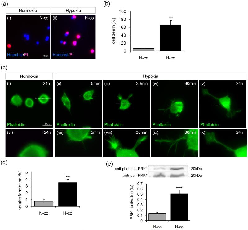
To induce hypoxia, PC12 cells were placed in a hypoxic incubator with a controlled atmosphere adjusted to 1% O2 and 5% CO2. Cell death was then evaluated by double staining with Hoechst and PI. Under hypoxia (6-8h) cell death (pink cells) in PC12 cells was significantly increased from 6.419% ± 0.327% (normoxic cells, N-co) to 66.159% ± 11.075% (hypoxic cells, H-co) (Fig. 1a and 1b). As shown in previous experiments, cell death further increased to 67.038% at 24h as opposed to 8h (data not shown; Podhraski et al. 2005). However, due to better evaluability of data regarding protective effects, the earlier time point was chosen for our studies. Hypoxic PC12 cells exhibited F-actin-rich microspikes (filopodia) and protrusion-like structures (lamellipodia) as early as 5-30 min after being switched to hypoxic conditions, leading to further neurite formation over a longer hypoxic incubation time [Fig. 1c (ii-v and vii-x)]. In contrast, normoxic control cells were largely unaffected, with smooth cell surfaces [Fig. 1c (i and vi)]. Quantification of neurite formation adequately showed a significant induction of neurite formation in PC12 cells (from 0.773% ± 0.218% under normoxia to 3.488% ± 0.537% under hypoxia) (Fig. 1d). PRK1 activity was increased in hypoxic cells after 5 min (0.507% ± 0.078%, compared to 0.137% ± 0.022% in normoxic cells) (Fig. 1e), and gradually ceased over time (6h 0.143% ± 0.041%; data not shown).
Fig. 1.
Hypoxia induces morphological changes and PRK1 activation in PC12 cells. PC12 cells were incubated under normoxic (21% O2; N-co) or hypoxic (1% O2; H-co) conditions. (a) After 6-8h incubation, cells were stained with Hoechst/PI for photomicrographs of viable (blue) and dead (pink) cells. (b) Quantification of the percentage of cell death [pink cells × 100/total cell number]. (c) F-actin–associated cytoskeletal reorganization over 0-24h normoxia/hypoxia in phalloidin-stained cells (green). Dashed lines in panels c(i-v) indicate the area selected for higher magnification in panels c(vi-x). (d) Neurite formation of PC12 cells after incubation under normoxic and hypoxic conditions for 24h. Three to five independent microscopic fields were studied and the percentage of neurite formation was calculated [neurite-bearing cells × 100/total cell number]. Extensions longer than twice the cell diameter were considered neurites. (e) Hypoxic treatment for 5 min increases PRK1 activation compared to normoxic cells. Cell lysates were analyzed by Western blot with anti-phospho-PRK1 and anti-pan-PRK1 antibodies. The intensity was scanned by a Personal Densitometer SI scanner (Molecular Dynamics) and the ratio (anti-phospho/anti-pan–PRK1) was calculated. Pictures and blots are representative of 3 to 5 independent experiments. Values represent the means ± S.E.M., n=3-5. Differences were analyzed using unpaired one-tailed t-test: (b) ++P<0.01, (d) ++P<0.01 and (e) +++P<0.001.
PRK1 is activated by the neuroprotective substances guanosine and nerve growth factor
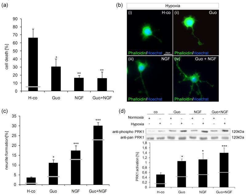
Next, the role of PRK1 in neuroprotective signaling was evaluated. Based on our own experience, we chose the purinergic substance guanosine and compared it to effects induced by NGF. The addition of ‘neurotrophic factors’ significantly decreased cell death of hypoxic PC12 cells from 66.159% ± 11.075% (H-co) to 30.159% ± 10.162% (guanosine), to 16.159% ± 3.531% (NGF) and to 15.3948% ± 7.916% (guanosine+NGF) (Fig. 2a). As mentioned above, staining of the F-actin cytoskeleton reveals characteristic rearrangements following incubation with low oxygen (H-co) [Fig. 2b (i)] that, remarkably, were supported by the addition of guanosine [Fig. 2b(ii)] or NGF [Fig. 2b (iii)]. Cells subjected to a combination of guanosine plus NGF formed the best-shaped neurites with growth-cones and branching [Fig. 2b(iv)]. Quantification of neurite formation revealed neurite formation under normoxia (white lines in columns): 4.012% ± 2.301% (guanosine), 12.829% ± 1.214% (NGF) and 22.635% ± 3.342% (guanosine+NGF). Under hypoxia, neurite formation was increased to 10.960% ± 1.746% (guanosine), 17.302% ± 2.624% (NGF), and to 29.963% ± 1.618% (guanosine plus NGF) (Fig. 2c). Upon addition of neurotrophins, PRK1 activity increased under normoxia (white lines in columns and representative blot) from 0.137% ± 0.022% (control) to 0.436% ± 0.100% (guanosine), 0.508% ± 0.163% (NGF), and 0.599% ± 0.188% (guanosine+NGF). Under hypoxic conditions, PRK1 activity increased from 0.507% ± 0.078% (control) to 1.054% ± 0.095% (guanosine), 1.119% ± 0.256 (NGF), and 1.388% ± 0.211 (guanosine+NGF) (Fig. 2d).
Fig. 2.
Guanosine and NGF mediate neuroprotective effects and increase PRK1 activation in PC12 cells. Hypoxic cells (H-co) were treated with 500μM guanosine (Guo), 5ng/mL NGF (NGF), or a combination of guanosine and NGF (Guo+NGF). (a) Cell death was quantified with Hoechst/PI after 6-8h treatment as described in experimental procedures. The white line indicates cell death in normoxic control. (b) F-actin–associated cytoskeleton was studied in phalloidin-stained cells (green) 24h after stimulation. Nuclei were labeled with Hoechst 33342 dye (blue). (c) Neurite formation was calculated in hypoxic PC12 cells (H-co), as well as in cells treated with 500μM guanosine (Guo), 5ng/mL NGF (NGF), or a combination of guanosine and NGF (Guo+NGF) for 24h. White lines indicate neurite formation under normoxic condition. (d) PRK1 activation was studied in cell lysates stimulated under normoxic/ hypoxic conditions and with 500μM guanosine (Guo), 5ng/mL NGF (NGF), or guanosine and NGF together (Guo+NGF) for 5 min by immunoprobing the blot with anti-phospho-PRK1 and anti-pan-PRK1 antibodies. White lines indicate PRK1 activation under normoxic condition. The intensity was scanned by a Personal Densitometer SI scanner (Molecular Dynamics) and the anti-phospho/anti-pan–PRK1 ratio was calculated. White lines indicate PRK1 activation under normoxic condition. Pictures and blots are representative of 4 to 11 independent experiments. Values represent the means ± S.E.M., n=3-11. Differences were analyzed using one-way ANOVA followed by Dunnett’s multiple comparison test: (a) *P<0.05, **P<0.01 (c) *P<0.05, ***P<0.001 and (d) *P<0.05, ***P<0.001.
PRK1 is essential for the rescue of hypoxic PC12 cells
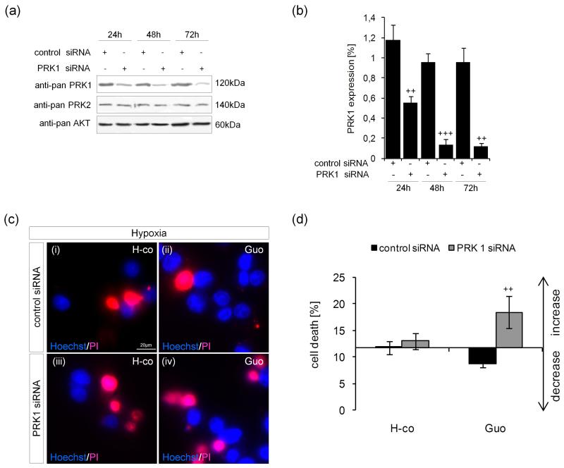
Cells were transfected with a synthetic siRNA construct directed against PRK1 or a scrambled, non-targeting control construct. Cell lysates were then analyzed for PRK1 and PRK2 expression by Western blot analysis. A remarkable downregulation of PRK1 expression was observed (Figs 3a and 3b). Specificity of PRK1 siRNA was confirmed by failure to change PRK2 expression levels (Fig. 3a). Twenty-four hours after transfection, a 52.97% reduction in PRK1 expression (from 1.180% ± 0.148% to 0.555% ± 0.061%) was observed. This reduction became greater over time: it was 85.88% (from 0.956% ± 0.085% to 0.135% ± 0.058%) at 48h and 87.62% (0.961% ± 0.141% to 0.119% ± 0.033%) at 72h (Fig. 3b). Accompanying PRK1 suppression, cell death increased in normoxic cultures by 49.09% in control cells and 76.06% in guanosine-treated cells (data not shown). Under hypoxic conditions, loss of functional PRK1 increased cell death by 10.49% in control cells (from 11.794% ± 1.198% in siRNA negative controls to 13.031% ± 1.497% in PRK knockdown cells) and by 110.27% in guanosine-treated cells (from 8.769% ± 0.743% in siRNA negative controls to 18.439% ± 2.971% in PRK knockdown cells) [Fig. 3c(i-iv) and 3d], illustrating the important role of PRK1 in purine-mediated neuroprotection of hypoxic PC12 cells.
Fig. 3.
siRNA-mediated PRK1 knockdown is specific and suppresses viability of hypoxic PC12 cells. PC12 cells were transfected with synthetic siRNA for PRK1 or scrambled non-targeting siRNA duplexes. (a) Cell extracts were analyzed 24h to 72h after transfection by immunoblotting for anti-pan-PRK1, anti-pan-PRK2 (specificity control) and anti-pan-AKT (loading control). (b) The intensity of protein bands was scanned by a Personal Densitometer SI scanner (Molecular Dynamics) and the anti-pan-PRK1/anti-pan-AKT ratio was calculated. (c) The effect of siRNA-mediated knockdown on cell viability was studied after additional stimulation of hypoxic PC12 cells (H-co) with 500μM guanosine (Guo) for 6-8h and staining for Hoechst/PI. (d) The percentage of hypoxic cell death in PRK1 knockdown cells was calculated. Pictures and blots are representative of 3 to 5 independent experiments and values represent the means ± S.E.M., n=3-5. Differences were analyzed using unpaired one-tailed t-test: (b) ++P<0.01, +++P<0.001 and (d) ++P<0.01 (control siRNA vs. PRK1 siRNA).
Effect of PRK1 knockdown on “neurotrophin”-mediated cell morphology, GAP-43 expression, and neurite formation in hypoxic PC12 cells
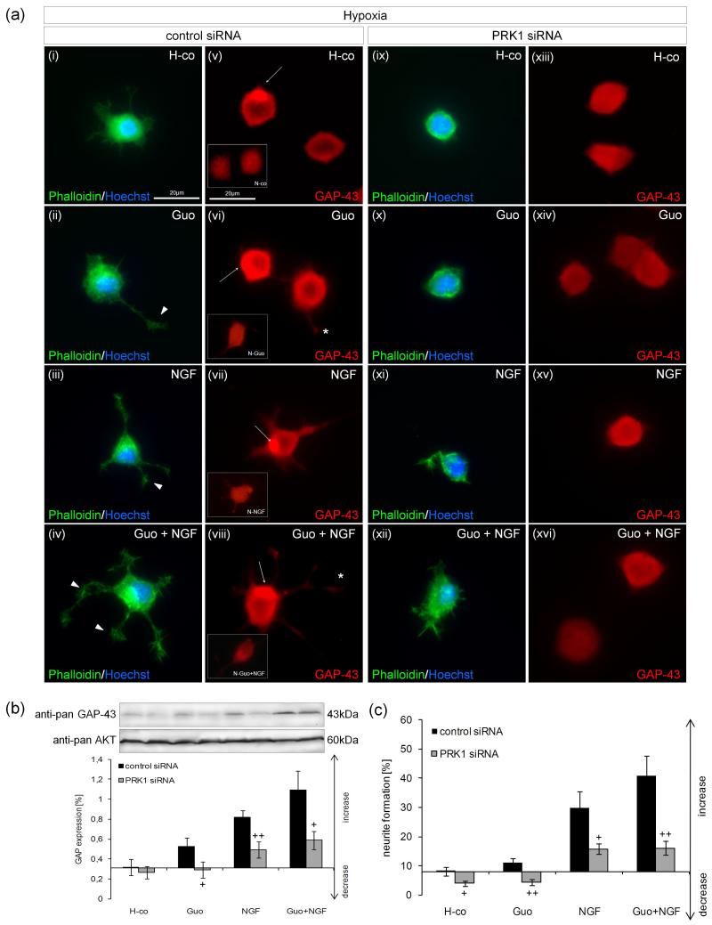
To clarify the question of whether PRK1 activation is essential to actin cytoskeletal regulation and subsequent neurite outgrowth, cells were transfected with siRNA constructs and incubated in 1% oxygen. In cells transfected with scrambled non-targeting control constructs, addition of the neurotrophic substances guanosine and NGF caused the formation of cytoplasmic extensions and accumulation of F-actin at the periphery of neurite growth cones and in branching points of neurites [arrowheads; Fig. 4a(ii-iv)]. However, upon siRNA-mediated knockdown of PRK1, F-actin-rich filopodia and growth cones were rarely formed, which indicates ineffective cytoskeletal reorganization [Fig. 4a(ix-xii)]. In parallel, cells were stained for the neuron-specific protein GAP-43 [Fig. 4a(v-viii and xiii-xvi)]. Normoxic cells lacking neurites demonstrated somal staining of GAP-43 [insert in Fig. 4a(v)]. Upon stimulation with guanosine and NGF a minor increase of GAP-43 staining was observed [inserts in Fig. 4a(vi-viii)]. Hypoxia induced the condensation of GAP-43 in ring-like structures, which were even more intense upon addition of neurotrophic substances [Fig. 4a(v-viii)]. Accompanying neurite formation, high levels of GAP-43 protein were observed in neurites and growth cones [asterisk; Fig. 4a(vi and viii)]. Furthermore, a perinuclear concentration of GAP-43 immunoreactivity was detected [arrows; Fig. 4a(v-viii)]. Knockdown of PRK1 caused a suppression of GAP-43 fluorescence intensity and disturbance of typical distribution patterns of GAP-43, as described above [Fig. 4a(xiii-xvi)]. Due to siRNA-mediated PRK knockdown, GAP-43 expression was inhibited in controls by 15.56% (from 0.315% ± 0.081% to 0.266% ± 0.063%), in guanosine-treated cultures by 45.16% (from 0.527% ± 0.086% to 0.289% ± 0.083%), in NGF-treated cultures by 39.46% (from 0.816% ± 0.068% to 0.494% ± 0.082%) and in cells treated with guanosine plus NGF by 46.29% (from 1.093% ± 0.190% to 0.587% ± 0.093%) (Fig. 4b).
Fig. 4.
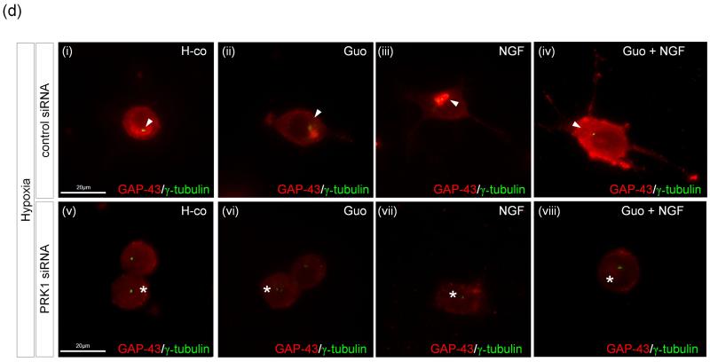
siRNA-mediated PRK1 knockdown inhibits guanosine- and NGF-mediated cytoskeletal reorganization, GAP-43 expression, and neurite formation in hypoxic PC12 cells. PC12 cells were transfected with siRNA for PRK1 or control siRNA duplexes. (a) siRNA-transfected hypoxic PC12 cells (H-co) were treated with 500μM guanosine (Guo), 5ng/mL NGF (NGF), or both guanosine and NGF (Guo+NGF) for 24h and then stained with FITC-phalloidin (green); nuclei were labeled with Hoechst 33342 (blue) [panels a(i-iv and ix-xii)]. F-actin accumulation at neurite growth cones and branching points was observed (arrowheads). In parallel, hypoxic neurons (H-co) were immunostained for anti-pan-GAP-43 (red) [panels a(v-viii and xiii-xvi), the inserts in panel a(v-viii) represent normoxic cells] after stimulation with 500μM guanosine (Guo), 5ng/mL NGF (NGF), or guanosine plus NGF (Guo+NGF) for 24h. Condensed levels of GAP-43 in growth cones (asterisk) and in perinuclei (arrows) are shown. Photomicrographs are representative of 3 to 5 independent experiments, each with 4 to 6 independent microscopic fields. (b) GAP-43 expression was investigated by analyzing cell lysates from transfected hypoxic PC12 cells (H-co) and hypoxic cells treated with 500μM guanosine (Guo), 50ng/mL NGF (NGF), or a combination of both (Guo+NGF) for 3 days. Intensity was scanned by a Personal Densitometer SI scanner (Molecular Dynamics) and the anti-pan-GAP-43/anti-pan-AKT ratio was calculated. (c) The effect of PRK1 knockdown on guanosine- and NGF-mediated neurite formation by stimulation of siRNA-transfected cells with 500μM guanosine (Guo), 5ng/mL NGF (NGF), or guanosine plus NGF (Guo+NGF) under hypoxic conditions for 3 days was calculated as described in experimental procedures. (d) Transfected PC12 cells treated with guanosine (Guo), 5ng/mL NGF (NGF), or guanosine plus NGF (Guo+NGF) under hypoxic conditions for 24h were stained for gamma tubulin (green) and anti-pan-GAP-43 (red) [panels d(i-viii)]. Pictures are representative of 3 independent experiments. Values represent the means ± S.E.M., n=3-9. Differences were analyzed using unpaired one-tailed t-test: (b) +P<0.05, ++P<0.01 and (c) +P<0.05, ++P<0.01 (control siRNA vs. PRK1 siRNA).
Analysis of neurite formation revealed analogous effects of PRK knockdown. Under hypoxic conditions, neurite formation was significantly reduced by siRNA-mediated PRK1 knockdown in controls by 51.69% (from 8.154% ± 1.471% to 3.939% ± 0.889%), in guanosine-treated cultures by 61.03% (from 11.151% ± 1.428% to 4.345% ± 1.052%), in NGF-treated cultures by 46.83% (from 29.717% ± 5.861% to 15.800% ± 1.737%) and in cells treated with guanosine plus NGF by 60.68% (from 40.964% ± 6.813% to 16.107% ± 2.312%) (Fig. 4c). Reduced neurotrophin-mediated neurite formation was also observed upon PRK1 knockdown in normoxic cultures: in controls by 40.93% (from 2.465% ± 0.391% to 1.456% ± 0.365%), in guanosine-treated cultures by 58.77% (from 5.168% ± 1.007% to 2.131% ± 0.256%), in NGF-treated cultures by 39.74% (from 28.515% ± 3.965% to 17.182% ± 1.670%) and in cells treated with guanosine plus NGF by 50.80% (from 36.354% ± 4.686% to 17.886% ± 1.091%) (data not shown). In neurons, the position of the centrosome marks the point of emergence of the future axon. Gamma tubulin staining of transfected PC12 cells showed that in control cells the centrosome indeed appears next to the newly formed neurite [arrowhead; Fig. 4d(i-iv)]; in comparison, the centrosome is not polarized upon siRNA-mediated PRK1 knockdown [asterisk; Fig. 4d(v-viii)]. Data therefore demonstrate the crucial role of PRK1 in the “neurotrophin”-mediated formation of neurite extensions.
Guanosine protects the viability and neuronal network of hypoxic primary cerebellar granule neurons
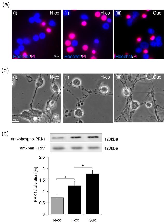
Similar to results obtained in PC12 cells, hypoxic insult disturbed the viability of primary neurons [Fig. 5a(i-ii)], but also hampered the neuronal network in cerebellar granule neuron cultures [Fig. 5b(i-ii)]. Additional incubation with guanosine potently rescued viability [Fig. 5a(iii)] and neurite formation in hypoxic cells [Fig. 5b(iii)]. In analogy to PC12 cells, subjection of cerebellar neurons to 1% oxygen again stimulated a significant increase in PRK1 activity by 72.51% (from 0.724% ± 0.157% to 1.249% ± 0.206%), which was enhanced by an additional 41.39% (from 1.249% ± 0.206% to 1.766% ± 0.183%) by the addition of guanosine (Fig. 5c).
Fig. 5.
Guanosine protects cell viability and the neuronal network in hypoxic primary cerebellar granule neurons. Primary cerebellar granule neurons were subjected to normoxia or hypoxia. (a) After 6-8h treatment normoxic (N-co) or hypoxic cells (H-co) were stained for viability with Hoechst/PI according to the protocol in experimental procedures [panels a(i and ii)]. Treatment with 500μM guanosine (Guo) reduced cell death [panels a(ii and iii)]. (b) In phase contrast photomicrographs, neurites of cerebellar granule neurons were visualized [panels b(i and ii)] after 6-8h incubation. Additional 500μM guanosine (Guo) improved stability of the neuronal network in hypoxic cerebellar granule cells [panels b(ii and iii)]. (c) PRK1 activation was studied after 5 min treatment under normoxic conditions (N-co), hypoxic conditions (H-co) and in hypoxic neurons incubated with 500μM guanosine (Guo). Pictures and blots are representative of 6 to 9 independent experiments. Values represent the means ± S.E.M., n=6-9. Differences were analyzed using unpaired one-tailed t-test: (c) +P<0.05.
PRK1 is essential for guanosine-mediated protection of hypoxic cerebellar granule neurons
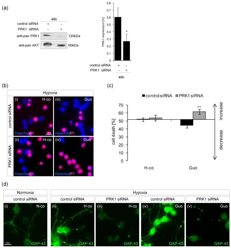
Cerebellar granule neurons were transfected with siRNA constructs directed against PRK1, whereupon expression was significantly and specifically suppressed from 0.603% ± 0.129% to 0.268% ± 0.101%, which equals a 55.56% downregulation (Fig. 6a). Under hypoxic conditions, PRK1 knockdown caused an increase in cell death in control cultures from 52.088% ± 2.241% to 53.728% ± 3.604%, which was not statistically significant. In guanosine-treated cultures, a significant increase in cell death (from 44.763% ± 3.091% to 61.658% ± 3.202%) was observed [Fig. 6b(i-iv) and c]. Normoxic cell cultures were also affected by PRK1 knockdown; cell death increased insignificantly in control cells from 48.730% ± 2.121% to 50.744% ± 2.292% and in guanosine-treated cells from 43.961% ± 3.451% to 53.433% ± 3.201% (data not shown).
Fig. 6.
PRK1 is essential for neuroprotection and GAP-43 expression in hypoxic cerebellar granule neurons. Freshly isolated primary cerebellar granule neurons were transfected with control or specific siRNA for PRK1. (a) PRK1 protein expression was studied 48h post-transfection by immunoblotting. The intensity was scanned by a Personal Densitometer SI scanner (Molecular Dynamics) and the anti-pan-PRK1/anti-pan-AKT ratio was calculated. (b) Transfected cerebellar granule neurons were exposed to hypoxic conditions (H-co) [panels b(i and ii)], treated with 500μM guanosine (Guo) [panels b(iii and iv)] for 6-8h, and stained with Hoechst/PI. (c) Quantification of the effect of PRK1 knockdown on cell death. (d) GAP-43 protein fluorescence intensity was studied by staining normoxic (N-co) [panel d(i)] and hypoxic cerebellar granule neurons (H-co) [panels d(ii and iii)]. Hypoxic cells were also treated with 500μM guanosine (Guo) [panels d(iv and v)]. Pictures and blots are representative of 3 to 4 independent experiments. Values represent the means ± S.E.M., n=4. Differences were analyzed using unpaired one-tailed t-test: (a) +P<0.05 and (c) ++P<0.01.
Next, GAP-43 fluorescence intensity was studied in transfected cerebellar granule neurons. Hypoxia-treated cells demonstrated an enhanced GAP-43 immunoreactivity compared to normoxic neurons [Fig. 6d(i-ii)], which was even exceeded by guanosine [Fig. 6d(iv)]. A striking downregulation of GAP-43 immunoreactivity was detected in PRK1-transfected cerebellar granule neurons [Fig. 6d(iii)] and even the addition of guanosine could not reverse this reduction [Fig. 6d(v)].
Discussion
As a consequence of an acute reduction of oxygen tension, several changes were noticed in PC12 cell cultures, including: a) increased cell death, b) reorganization of the cytoskeleton associated with the formation of characteristic F-actin-rich filopodia and lamellipodia after 5-30 min in hypoxic conditions, leading, upon prolonged hypoxia, to c) neurite formation. In analogy to earlier reports (Tomaselli et al. 2008; Zur Nedden et al. 2008), primary cerebellar granule neurons were also considerably compromised by a low oxygen atmosphere. Concomitant with morphological changes after culture in low oxygen for 5 min (F-actin-rich filopodia and lamellipodia), a significant increase in the activity of the protein kinase C-related kinase PRK1 was induced. PRK1 is a PKC family member that is involved in the regulation of intermediate filaments of the actin cytoskeleton, as well as in processes as diverse as mitotic timing and apoptosis (for review see Modha et al. 2008). In support of our findings, others have previously observed that PRK1 is regulated by hyperosmotic shock (Torbett et al. 2003), and that stress in general leads to an increased nuclear translocation of PRK (Mukai et al. 1996). Along this line, an increased nuclear fraction of PRK1 was recently also detected in hypoxic PC12 cells (S. z. Nedden, unpublished data). Based on our previous work on neuronal differentiation and the stress response in PC12 cells and cerebellar granule neurons, the remarkable neurite-inducing capability of guanosine (Bocklinger et al. 2004; Heftberger et al. 2005; Tomaselli et al. 2005a; Tomaselli et al. 2005b; Tomaselli et al. 2008), and an indication that PRK activation may be involved in purine-mediated signaling (Tomaselli et al. 2005a), we wanted to clarify whether PRK1 plays a specific role in the neuroprotection of hypoxic cells that is mediated by guanosine and by NGF.
As a first step, hypoxic cells were incubated with guanosine, NGF, or a combination of the two. Our results revealed that guanosine and NGF not only potently rescued hypoxic PC12 cells and cerebellar granule neurons, but also significantly enhanced hypoxia-induced PRK activity in these cells. The combination of guanosine and NGF at most led to an additive enhancement, impying that the pathways involved overlap at least partially. A strong link between purine nucleosides and hypoxia-related signaling already exists (for reviews, see Fredholm 2007; Sitkovsky et al. 2008;Tomaselli et al. 2008; and Zur Nedden et al. 2008). The expression levels of purine nucleosides and adenosine receptors are regulated in conditions of cellular stress. Hypoxia apparently induces an expression program that shifts the tissue phenotype toward an increase in extracellular purine nucleosides. In turn, adenosine receptor activation tends to limit the potential damage incurred by hypoxia (Fredholm 2007). Whether the protective effect of guanosine is at least partly mediated by adenosine or its receptors (Ciccarelli et al. 2000; D’Alimonte et al. 2007), or is mediated by its own specific G-coupled receptors (Traversa et al. 2003), is still controversial. Our own data suggest at least a supporting role for the A2A receptor in guanosine-mediated signal transduction in neurite formation (B. Thauerer, unpublished data). Regarding data by others (Oleskovicz et al. 2008), guanosine may activate a receptor or a calcium-dependent K+ channel, promoting PKC, MEK, PI3-K, or PKA activation, which have been directly associated with neuroprotective mechanisms against ischemic damage (Love 2003; Horn et al. 2005). Likewise, TrkA-receptor-mediated NGF signaling apparently also leads to PKA activation, which in turn induces RhoA phosphorylation at serine 188 in the C-terminus (Nusser et al. 2002). Phosphorylation of this site then selectively inhibits interaction between RhoA and ROCK (Rho kinase), but enhances complex formation of RhoA with PRK. Neuritogenesis is one of the first steps of neuronal differentiation in which the RhoA/ROCK system plays a decisive role (Govek et al. 2005). Activation of RhoA induces neurite retraction in neuronal cell lines. Conversely, inactivation of RhoA or its downstream effectors, the ROCKs, enhances neurite extension and growth cone movement (Hirose et al. 1998; Bito et al. 2000). Presently, two ROCK isoforms have been identified, ROCK1 and ROCK2, whose activity can be inhibited with the ROCK inhibitor Y-27632 (Ishizaki et al. 2000). Our own data (B. Thauerer, unpublished data) revealed that upon incubation with Y-27632, PC12 cells develop significantly more neurites than control cultures do. To further study the non-redundant role of PRK in the rescue of hypoxic PC12 cells and cerebellar granule neurons, we next employed an siRNA approach in order to specifically suppress PRK1 activation. Following siRNA-mediated knockdown of PRK1 expression, we observed decreased viability and neurite formation in PC12 cells and cerebellar granule neurons. Concomitantly, siRNA-mediated PRK1 knockdown deterred guanosine- and NGF-mediated F-actin rearrangement and associated neurite formation, and completely inhibited purine nucleoside-mediated protective effects. However, due to the different stages of neuronal polarization in our two cell models (in contrast to PC12 cells, p7 cerebellar granule neurons are more differentiated) comparability of the results from these two models is limited. Current findings, as well as other reports showing an involvement of PRK1 in the regulation of the actin cytoskeleton (for review see Sun et al. 2000), supported our hypothesis that PRK1 activation is essential for neuronal plasticity.
The next challenge was to link this substantial finding with other molecular or genetic events, and eventually with physiological mechanisms. According to earlier reports, NGF-mediated differentiation of PC12 cells leads to increased immunoreactivity of GAP-43 (Costello et al. 1990; O’Driscoll and Gorman 2005) due to protein kinase C-dependent stabilization of the GAP-43 mRNA (Perrone-Bizzozero et al. 1993; Irwin et al. 2002). GAP-43 is a neuron-specific protein that exhibits elevated synthesis and axonal fast transport during development and nerve regeneration (Benowitz et al. 1981; Snipes et al. 1987; for a review see Skene 1989). Others (Perrone-Bizzozero et al. 1986) observed increased GAP-43 expression in cultured neurons when maintained under conditions promoting neurite outgrowth. We also observed increased GAP-43 immunoreactivity in hypoxic PC12 cells and cerebellar granule neurons compared to normoxic controls. Incubation with NGF, and even more remarkably with guanosine, further significantly enhanced GAP-43 expression in PC12 cells. Apparently, GAP-43 protein accumulates at the plasma membrane; consequently, the effect of PRK1 knockdown on GAP-43 expression was studied. To verify this connection, PC12 cells and cerebellar granule cells were transfected with siRNA constructs targeting PRK1. As a result we detected considerable suppression of GAP-43 immunoreactivity in PC12 cells and cerebellar granule neurons, as well as GAP-43 expression in PC12 cells. In neurons, centrosomal position marks the point of emergence of the future axon. As previously shown by others (Mishra et al. 2008), GAP-43 is key to mitotic spindle control and centrosome-based polarization in neurons. Here, gamma tubulin staining of transfected PC12 cells demonstrated that while the centrosome appeared to localize next to the newly formed neurite in control cells, the centrosome was apparently no longer polarized upon siRNA-mediated PRK1 knockdown (Fig. 4d).
In conclusion, our results have confirmed earlier findings by Tomaselli et al. 2008 and Zur Nedden et al. 2008, suggesting that hypoxic treatment stimulates cell death and neurite formation. We have also demonstrated that hypoxic conditions concomitantly increase PRK1 activity as monitored by autophosphorylation. In addition, our data indicate that guanosine is neuroprotective in the presence of hypoxia, and stimulates neurite outgrowth and PRK1 activity. The combination of guanosine and NGF led to an additive enhancement of NGF-mediated neurite outgrowth. However, guanosine did not significantly amplify NGF-induced neuroprotection or NGF-mediated PRK1 activation, implicating the involvement of at least partially overlapping pathways. Our findings also suggest that when PRK1 is downregulated with siRNA, both guanosine-stimulated neuroprotection and neurite outgrowth are inhibited, and GAP-43 is downregulated. Finally, guanosine has the same effects on hypoxia-mediated cell viability and GAP-43 downregulation in cerebellar granule neurons, and stabilizes existing assaulted neurites in this culture system.
Brain exposure to hypoxia in ischemia/reperfusion injuries often causes devastating and irreversible losses of function (Chen et al. 2002) and is linked to long term neurological deficits (Berger and Garnier 1999; Davis et al. 2000; El-Khodor and Boksa 2000; Boksa and El-Khodor 2003). Knowing how regeneration occurs may allow novel innovations in the protection of the nervous system. The detection of a substantial role for PRK1 in neuroprotection and regeneration should therefore support current efforts to exploit signaling mechanisms for the development of neuroprotective pharmacological substances.
Acknowledgements
This work was supported by a grant of the Austrian FWF P19578-B05. We are grateful to Dr. G. Baier and Dr. C. Bandtlow for helpful discussion and support.
Abbreviations used
- A2AR
A2A adenosine receptor
- BSA
bovine serum albumin
- GAP-43
growth associated protein-43
- NGF
nerve growth factor
- PBS
phosphate buffered saline
- PI
propidium iodide
- PKC
protein kinase C
- PRK1
protein kinase C-related kinase
- RT
room temperature
- siRNA
small interfering RNA
- PC12
pheochromocytoma cells
References
- Arslan G, Kull B, Fredholm BB. Signaling via A2A adenosine receptor in four PC12 cell clones. Naunyn Schmiedebergs Arch Pharmacol. 1999;359:28–32. doi: 10.1007/pl00005319. [DOI] [PubMed] [Google Scholar]
- Benowitz LI, Shashoua VE, Yoon MG. Specific changes in rapidly transported proteins during regeneration of the goldfish optic nerve. J Neurosci. 1981;1:300–307. doi: 10.1523/JNEUROSCI.01-03-00300.1981. [DOI] [PMC free article] [PubMed] [Google Scholar]
- Berger R, Garnier Y. Pathophysiology of perinatal brain damage. Brain Res Brain Res Rev. 1999;30:107–134. doi: 10.1016/s0165-0173(99)00009-0. [DOI] [PubMed] [Google Scholar]
- Bito H, Furuyashiki T, Ishihara H, Shibasaki Y, Ohashi K, Mizuno K, Maekawa M, Ishizaki T, Narumiya S. A critical role for a Rho-associated kinase, p160ROCK, in determining axon outgrowth in mammalian CNS neurons. Neuron. 2000;26:431–441. doi: 10.1016/s0896-6273(00)81175-7. [DOI] [PubMed] [Google Scholar]
- Bocklinger K, Tomaselli B, Heftberger V, Podhraski V, Bandtlow C, Baier-Bitterlich G. Purine nucleosides support the neurite outgrowth of primary rat cerebellar granule cells after hypoxia. Eur J Cell Biol. 2004;83:51–54. doi: 10.1078/0171-9335-00362. [DOI] [PubMed] [Google Scholar]
- Boksa P, El-Khodor BF. Birth insult interacts with stress at adulthood to alter dopaminergic function in animal models: possible implications for schizophrenia and other disorders. Neurosci Biobehav Rev. 2003;27:91–101. doi: 10.1016/s0149-7634(03)00012-5. [DOI] [PubMed] [Google Scholar]
- Chen P, Goldberg DE, Kolb B, Lanser M, Benowitz LI. Inosine induces axonal rewiring and improves behavioral outcome after stroke. Proc Natl Acad Sci U S A. 2002;99:9031–9036. doi: 10.1073/pnas.132076299. [DOI] [PMC free article] [PubMed] [Google Scholar]
- Ciccarelli R, Di Iorio P, D’Alimonte I, Giuliani P, Florio T, Caciagli F, Middlemiss PJ, Rathbone MP. Cultured astrocyte proliferation induced by extracellular guanosine involves endogenous adenosine and is raised by the co-presence of microglia. Glia. 2000;29:202–211. [PubMed] [Google Scholar]
- Costello B, Meymandi A, Freeman JA. Factors influencing GAP-43 gene expression in PC12 pheochromocytoma cells. J Neurosci. 1990;10:1398–1406. doi: 10.1523/JNEUROSCI.10-04-01398.1990. [DOI] [PMC free article] [PubMed] [Google Scholar]
- D’Alimonte I, Flati V, D’Auro M, Toniato E, Martinotti S, Rathbone MP, Jiang S, Ballerini P, Di Iorio P, Caciagli F, Ciccarelli R. Guanosine inhibits CD40 receptor expression and function induced by cytokines and beta amyloid in mouse microglia cells. J Immunol. 2007;178:720–731. doi: 10.4049/jimmunol.178.2.720. [DOI] [PubMed] [Google Scholar]
- Davis S, Vanhoutte P, Pages C, Caboche J, Laroche S. The MAPK/ERK cascade targets both Elk-1 and cAMP response element-binding protein to control long-term potentiation-dependent gene expression in the dentate gyrus in vivo. J Neurosci. 2000;20:4563–4572. doi: 10.1523/JNEUROSCI.20-12-04563.2000. [DOI] [PMC free article] [PubMed] [Google Scholar]
- El-Khodor BF, Boksa P. Transient birth hypoxia increases behavioral responses to repeated stress in the adult rat. Behav Brain Res. 2000;107:171–175. doi: 10.1016/s0166-4328(99)00119-9. [DOI] [PubMed] [Google Scholar]
- Fredholm BB. Adenosine, an endogenous distress signal, modulates tissue damage and repair. Cell Death Differ. 2007;14:1315–1323. doi: 10.1038/sj.cdd.4402132. [DOI] [PubMed] [Google Scholar]
- Gartner A, Collin L, Lalli G. Nucleofection of primary neurons. Methods Enzymol. 2006;406:374–388. doi: 10.1016/S0076-6879(06)06027-7. [DOI] [PubMed] [Google Scholar]
- Govek EE, Newey SE, Van Aelst L. The role of the Rho GTPases in neuronal development. Genes Dev. 2005;19:1–49. doi: 10.1101/gad.1256405. [DOI] [PubMed] [Google Scholar]
- Greene LA, Tischler AS. Establishment of a noradrenergic clonal line of rat adrenal pheochromocytoma cells which respond to nerve growth factor. Proc Natl Acad Sci U S A. 1976;73:2424–2428. doi: 10.1073/pnas.73.7.2424. [DOI] [PMC free article] [PubMed] [Google Scholar]
- Gudi T, Chen JC, Casteel DE, Seasholtz TM, Boss GR, Pilz RB. cGMP-dependent protein kinase inhibits serum-response element-dependent transcription by inhibiting rho activation and functions. J Biol Chem. 2002;277:37382–37393. doi: 10.1074/jbc.M204491200. [DOI] [PubMed] [Google Scholar]
- Gysbers JW, Rathbone MP. Neurite outgrowth in PC12 cells is enhanced by guanosine through both cAMP-dependent and -independent mechanisms. Neurosci Lett. 1996;220:175–178. doi: 10.1016/s0304-3940(96)13253-5. [DOI] [PubMed] [Google Scholar]
- Hatten ME. Neuronal regulation of astroglial morphology and proliferation in vitro. J Cell Biol. 1985;100:384–396. doi: 10.1083/jcb.100.2.384. [DOI] [PMC free article] [PubMed] [Google Scholar]
- Hatten ME, Alder J, Zimmerman K, Heintz N. Genes involved in cerebellar cell specification and differentiation. Curr Opin Neurobiol. 1997;7:40–47. doi: 10.1016/s0959-4388(97)80118-3. [DOI] [PubMed] [Google Scholar]
- Heftberger V, Tomaselli B, Podhraski V, Baier-Bitterlich G. Purine Nucleoside Mediated Protection of Primary Cerebellar Granule Cells after Hypoxic Insult. Nova Science Publishers, Inc; New York: 2005. [Google Scholar]
- Hide I, Padgett WL, Jacobson KA, Daly JW. A2A adenosine receptors from rat striatum and rat pheochromocytoma PC12 cells: characterization with radioligand binding and by activation of adenylate cyclase. Mol Pharmacol. 1992;41:352–359. [PMC free article] [PubMed] [Google Scholar]
- Hirose M, Ishizaki T, Watanabe N, Uehata M, Kranenburg O, Moolenaar WH, Matsumura F, Maekawa M, Bito H, Narumiya S. Molecular dissection of the Rho-associated protein kinase (p160ROCK)-regulated neurite remodeling in neuroblastoma N1E-115 cells. J Cell Biol. 1998;141:1625–1636. doi: 10.1083/jcb.141.7.1625. [DOI] [PMC free article] [PubMed] [Google Scholar]
- Horn AP, Gerhardt D, Geyer AB, Valentim L, Cimarosti H, Tavares A, Horn F, Lenz G, Salbego C. Cellular death in hippocampus in response to PI3K pathway inhibition and oxygen and glucose deprivation. Neurochem Res. 2005;30:355–361. doi: 10.1007/s11064-005-2609-0. [DOI] [PubMed] [Google Scholar]
- Irwin N, Chao S, Goritchenko L, Horiuchi A, Greengard P, Nairn AC, Benowitz LI. Nerve growth factor controls GAP-43 mRNA stability via the phosphoprotein ARPP-19. Proc Natl Acad Sci U S A. 2002;99:12427–12431. doi: 10.1073/pnas.152457399. [DOI] [PMC free article] [PubMed] [Google Scholar]
- Ishizaki T, Uehata M, Tamechika I, Keel J, Nonomura K, Maekawa M, Narumiya S. Pharmacological properties of Y-27632, a specific inhibitor of rho-associated kinases. Mol Pharmacol. 2000;57:976–983. [PubMed] [Google Scholar]
- Kobayashi S, Millhorn DE. Stimulation of expression for the adenosine A2A receptor gene by hypoxia in PC12 cells. A potential role in cell protection. J Biol Chem. 1999;274:20358–20365. doi: 10.1074/jbc.274.29.20358. [DOI] [PubMed] [Google Scholar]
- Kobayashi S, Conforti L, Pun RY, Millhorn DE. Adenosine modulates hypoxia-induced responses in rat PC12 cells via the A2A receptor. J Physiol. 1998;508(Pt 1):95–107. doi: 10.1111/j.1469-7793.1998.095br.x. [DOI] [PMC free article] [PubMed] [Google Scholar]
- Kuhar SG, Feng L, Vidan S, Ross ME, Hatten ME, Heintz N. Changing patterns of gene expression define four stages of cerebellar granule neuron differentiation. Development. 1993;117:97–104. doi: 10.1242/dev.117.1.97. [DOI] [PubMed] [Google Scholar]
- Love S. Apoptosis and brain ischaemia. Prog Neuropsychopharmacol Biol Psychiatry. 2003;27:267–282. doi: 10.1016/S0278-5846(03)00022-8. [DOI] [PubMed] [Google Scholar]
- Lu Y, Settleman J. The Drosophila Pkn protein kinase is a Rho/Rac effector target required for dorsal closure during embryogenesis. Genes Dev. 1999;13:1168–1180. doi: 10.1101/gad.13.9.1168. [DOI] [PMC free article] [PubMed] [Google Scholar]
- Mellor H, Parker PJ. The extended protein kinase C superfamily. Biochem J. 1998;332(Pt 2):281–292. doi: 10.1042/bj3320281. [DOI] [PMC free article] [PubMed] [Google Scholar]
- Mishra R, Gupta SK, Meiri KF, Fong M, Thostrup P, Juncker D, Mani S. GAP-43 is key to mitotic spindle control and centrosome-based polarization in neurons. Cell Cycle. 2008;7:348–357. doi: 10.4161/cc.7.3.5235. [DOI] [PubMed] [Google Scholar]
- Modha R, Campbell LJ, Nietlispach D, Buhecha HR, Owen D, Mott HR. The Rac1 polybasic region is required for interaction with its effector PRK1. J Biol Chem. 2008;283:1492–1500. doi: 10.1074/jbc.M706760200. [DOI] [PubMed] [Google Scholar]
- Mukai H. The structure and function of PKN, a protein kinase having a catalytic domain homologous to that of PKC. J Biochem (Tokyo) 2003;133:17–27. doi: 10.1093/jb/mvg019. [DOI] [PubMed] [Google Scholar]
- Mukai H, Miyahara M, Sunakawa H, Shibata H, Toshimori M, Kitagawa M, Shimakawa M, Takanaga H, Ono Y. Translocation of PKN from the cytosol to the nucleus induced by stresses. Proc Natl Acad Sci U S A. 1996;93:10195–10199. doi: 10.1073/pnas.93.19.10195. [DOI] [PMC free article] [PubMed] [Google Scholar]
- Neary JT, Rathbone MP, Cattabeni F, Abbracchio MP, Burnstock G. Trophic actions of extracellular nucleotides and nucleosides on glial and neuronal cells. Trends Neurosci. 1996;19:13–18. doi: 10.1016/0166-2236(96)81861-3. [DOI] [PubMed] [Google Scholar]
- Nusser N, Gosmanova E, Zheng Y, Tigyi G. Nerve growth factor signals through TrkA, phosphatidylinositol 3-kinase, and Rac1 to inactivate RhoA during the initiation of neuronal differentiation of PC12 cells. J Biol Chem. 2002;277:35840–35846. doi: 10.1074/jbc.M203617200. [DOI] [PubMed] [Google Scholar]
- O’Driscoll CM, Gorman AM. Hypoxia induces neurite outgrowth in PC12 cells that is mediated through adenosine A2A receptors. Neuroscience. 2005;131:321–329. doi: 10.1016/j.neuroscience.2004.11.015. [DOI] [PubMed] [Google Scholar]
- Oleskovicz SP, Martins WC, Leal RB, Tasca CI. Mechanism of guanosine-induced neuroprotection in rat hippocampal slices submitted to oxygen-glucose deprivation. Neurochem Int. 2008;52:411–418. doi: 10.1016/j.neuint.2007.07.017. [DOI] [PubMed] [Google Scholar]
- Perrone-Bizzozero NI, Finklestein SP, Benowitz LI. Synthesis of a growth-associated protein by embryonic rat cerebrocortical neurons in vitro. J Neurosci. 1986;6:3721–3730. doi: 10.1523/JNEUROSCI.06-12-03721.1986. [DOI] [PMC free article] [PubMed] [Google Scholar]
- Perrone-Bizzozero NI, Cansino VV, Kohn DT. Posttranscriptional regulation of GAP-43 gene expression in PC12 cells through protein kinase C-dependent stabilization of the mRNA. J Cell Biol. 1993;120:1263–1270. doi: 10.1083/jcb.120.5.1263. [DOI] [PMC free article] [PubMed] [Google Scholar]
- Podhraski V, Tomaselli B, Baier-Bitterlich G. Adenosine receptor expression in PC12-cells during normoxic and hypoxic conditions. J Neurochem. 2005;(Suppl 2):210–266. [Google Scholar]
- Rathbone MP, Middlemiss P, Andrew C, Caciagli F, Ciccarelli R, Di Iorio P, Huang R. The trophic effects of purines and purinergic signaling in pathologic reactions of astrocytes. Alzheimer Dis Assoc Disord. 1998;12(Suppl 2):S36–45. doi: 10.1097/00002093-199803001-00006. [DOI] [PubMed] [Google Scholar]
- Schmidt AP, Lara DR, Souza DO. Proposal of a guanine-based purinergic system in the mammalian central nervous system. Pharmacol Ther. 2007;116:401–416. doi: 10.1016/j.pharmthera.2007.07.004. [DOI] [PubMed] [Google Scholar]
- Seta KA, Spicer Z, Yuan Y, Lu G, Millhorn DE. Responding to hypoxia: lessons from a model cell line. Sci STKE 2002. 2002:RE11. doi: 10.1126/stke.2002.146.re11. [DOI] [PubMed] [Google Scholar]
- Sitkovsky MV, Kjaergaard J, Lukashev D, Ohta A. Hypoxia-adenosinergic immunosuppression: tumor protection by T regulatory cells and cancerous tissue hypoxia. Clin Cancer Res. 2008;14:5947–5952. doi: 10.1158/1078-0432.CCR-08-0229. [DOI] [PubMed] [Google Scholar]
- Skene JH. Axonal growth-associated proteins. Annu Rev Neurosci. 1989;12:127–156. doi: 10.1146/annurev.ne.12.030189.001015. [DOI] [PubMed] [Google Scholar]
- Snipes GJ, Costello B, McGuire CB, Mayes BN, Bock SS, Norden JJ, Freeman JA. Regulation of specific neuronal and nonneuronal proteins during development and following injury in the rat central nervous system. Prog Brain Res. 1987;71:155–175. doi: 10.1016/s0079-6123(08)61821-x. [DOI] [PubMed] [Google Scholar]
- Sun W, Vincent S, Settleman J, Johnson GL. MEK kinase 2 binds and activates protein kinase C-related kinase 2. Bifurcation of kinase regulatory pathways at the level of an MAPK kinase kinase. J Biol Chem. 2000;275:24421–24428. doi: 10.1074/jbc.M003148200. [DOI] [PubMed] [Google Scholar]
- Takahashi M, Mukai H, Toshimori M, Miyamoto M, Ono Y. Proteolytic activation of PKN by caspase-3 or related protease during apoptosis. Proc Natl Acad Sci U S A. 1998;95:11566–11571. doi: 10.1073/pnas.95.20.11566. [DOI] [PMC free article] [PubMed] [Google Scholar]
- Thangnipon W, Kingsbury A, Webb M, Balazs R. Observations on rat cerebellar cells in vitro: influence of substratum, potassium concentration and relationship between neurones and astrocytes. Brain Res. 1983;313:177–189. doi: 10.1016/0165-3806(83)90215-8. [DOI] [PubMed] [Google Scholar]
- Tomaselli B, Podhraski V, Böck G, Baier-Bitterlich G. Early cellular responses of purine nucleoside-mediated protection of hypoxia-induced injuries of neuronal PC12 cells. American Journal of Biochemistry and Biotechnology. 2005a;2:161–167. [Google Scholar]
- Tomaselli B, Nedden SZ, Podhraski V, Baier-Bitterlich G. p42/44 MAPK is an essential effector for purine nucleoside-mediated neuroprotection of hypoxic PC12 cells and primary cerebellar granule neurons. Mol Cell Neurosci. 2008;38:559–568. doi: 10.1016/j.mcn.2008.05.004. [DOI] [PubMed] [Google Scholar]
- Tomaselli B, Podhraski V, Heftberger V, Bock G, Baier-Bitterlich G. Purine nucleoside-mediated protection of chemical hypoxia-induced neuronal injuries involves p42/44 MAPK activation. Neurochem Int. 2005b;46:513–521. doi: 10.1016/j.neuint.2005.02.003. [DOI] [PubMed] [Google Scholar]
- Torbett NE, Casamassima A, Parker PJ. Hyperosmotic-induced protein kinase N 1 activation in a vesicular compartment is dependent upon Rac1 and 3-phosphoinositide-dependent kinase 1. J Biol Chem. 2003;278:32344–32351. doi: 10.1074/jbc.M303532200. [DOI] [PubMed] [Google Scholar]
- Traversa U, Bombi G, Camaioni E, Macchiarulo A, Costantino G, Palmieri C, Caciagli F, Pellicciari R. Rat brain guanosine binding site. Biological studies and pseudo-receptor construction. Bioorg Med Chem. 2003;11:5417–5425. doi: 10.1016/j.bmc.2003.09.043. [DOI] [PubMed] [Google Scholar]
- Uemura Y, Miller JM, Matson WR, Beal MF. Neurochemical analysis of focal ischemia in rats. Stroke. 1991;22:1548–1553. doi: 10.1161/01.str.22.12.1548. [DOI] [PubMed] [Google Scholar]
- van der Ploeg I, Ahlberg S, Parkinson FE, Olsson RA, Fredholm BB. Functional characterization of adenosine A2 receptors in Jurkat cells and PC12 cells using adenosine receptor agonists. Naunyn Schmiedebergs Arch Pharmacol. 1996;353:250–260. doi: 10.1007/BF00168626. [DOI] [PubMed] [Google Scholar]
- Velier JJ, Ellison JA, Kikly KK, Spera PA, Barone FC, Feuerstein GZ. Caspase-8 and caspase-3 are expressed by different populations of cortical neurons undergoing delayed cell death after focal stroke in the rat. J Neurosci. 1999;19:5932–5941. doi: 10.1523/JNEUROSCI.19-14-05932.1999. [DOI] [PMC free article] [PubMed] [Google Scholar]
- Vincent S, Settleman J. The PRK2 kinase is a potential effector target of both Rho and Rac GTPases and regulates actin cytoskeletal organization. Mol Cell Biol. 1997;17:2247–2256. doi: 10.1128/mcb.17.4.2247. [DOI] [PMC free article] [PubMed] [Google Scholar]
- Zetterstrom T, Vernet L, Ungerstedt U, Tossman U, Jonzon B, Fredholm BB. Purine levels in the intact rat brain. Studies with an implanted perfused hollow fibre. Neurosci Lett. 1982;29:111–115. doi: 10.1016/0304-3940(82)90338-x. [DOI] [PubMed] [Google Scholar]
- Zhu WH, Conforti L, Czyzyk-Krzeska MF, Millhorn DE. Membrane depolarization in PC-12 cells during hypoxia is regulated by an O2-sensitive K+ current. Am J Physiol. 1996;271:C658–665. doi: 10.1152/ajpcell.1996.271.2.C658. [DOI] [PubMed] [Google Scholar]
- Zur Nedden S, Tomaselli B, Baier-Bitterlich G. HIF-1 alpha is an essential effector for purine nucleoside-mediated neuroprotection against hypoxia in PC12 cells and primary cerebellar granule neurons. J Neurochem. 2008 doi: 10.1111/j.1471-4159.2008.05275.x. [DOI] [PMC free article] [PubMed] [Google Scholar]


