Fig. 5.
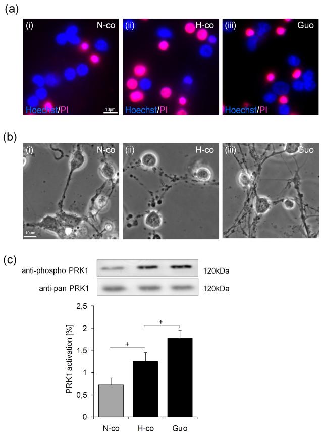
Guanosine protects cell viability and the neuronal network in hypoxic primary cerebellar granule neurons. Primary cerebellar granule neurons were subjected to normoxia or hypoxia. (a) After 6-8h treatment normoxic (N-co) or hypoxic cells (H-co) were stained for viability with Hoechst/PI according to the protocol in experimental procedures [panels a(i and ii)]. Treatment with 500μM guanosine (Guo) reduced cell death [panels a(ii and iii)]. (b) In phase contrast photomicrographs, neurites of cerebellar granule neurons were visualized [panels b(i and ii)] after 6-8h incubation. Additional 500μM guanosine (Guo) improved stability of the neuronal network in hypoxic cerebellar granule cells [panels b(ii and iii)]. (c) PRK1 activation was studied after 5 min treatment under normoxic conditions (N-co), hypoxic conditions (H-co) and in hypoxic neurons incubated with 500μM guanosine (Guo). Pictures and blots are representative of 6 to 9 independent experiments. Values represent the means ± S.E.M., n=6-9. Differences were analyzed using unpaired one-tailed t-test: (c) +P<0.05.

