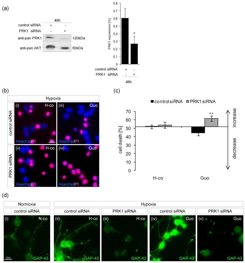
Fig. 6.
PRK1 is essential for neuroprotection and GAP-43 expression in hypoxic cerebellar granule neurons. Freshly isolated primary cerebellar granule neurons were transfected with control or specific siRNA for PRK1. (a) PRK1 protein expression was studied 48h post-transfection by immunoblotting. The intensity was scanned by a Personal Densitometer SI scanner (Molecular Dynamics) and the anti-pan-PRK1/anti-pan-AKT ratio was calculated. (b) Transfected cerebellar granule neurons were exposed to hypoxic conditions (H-co) [panels b(i and ii)], treated with 500μM guanosine (Guo) [panels b(iii and iv)] for 6-8h, and stained with Hoechst/PI. (c) Quantification of the effect of PRK1 knockdown on cell death. (d) GAP-43 protein fluorescence intensity was studied by staining normoxic (N-co) [panel d(i)] and hypoxic cerebellar granule neurons (H-co) [panels d(ii and iii)]. Hypoxic cells were also treated with 500μM guanosine (Guo) [panels d(iv and v)]. Pictures and blots are representative of 3 to 4 independent experiments. Values represent the means ± S.E.M., n=4. Differences were analyzed using unpaired one-tailed t-test: (a) +P<0.05 and (c) ++P<0.01.

