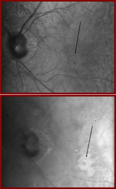
Figure 11.
Retinal images acquired with a Ti:Sapphire laser of a patient with a new blood vessel membrane indicated by black arrows,. The left image was acquired as in ref 26, at 860 nm and using a confocal aperture, showing only the superficial part of the new vessel membrane. The right image was acquired with an annular aperture, and the extent of the new blood vessel membrane is clearly larger than that indicated by the superficial information.

