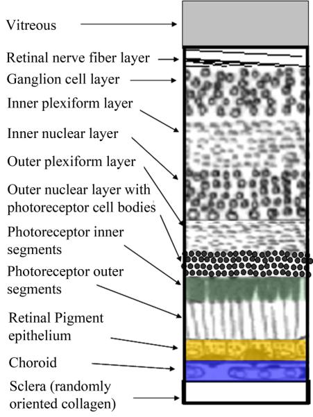
Figure 2.
Schematic of the layers of the retina, with the most superficial at the top. The retinal pigment epithelium is shown in gold, to correspond to its most prominent fluorophore, lipofucsin. The choroid is shown in blue, to correspond to its absorption in the short wavelength range due to melanin, which is strong in darkly pigmented eyes. The photoreceptor outer segments are shown in green, to correspond to their broad band absorption of the three most common photopigments, peaking roughly at 500, 535, and 565 nm.

