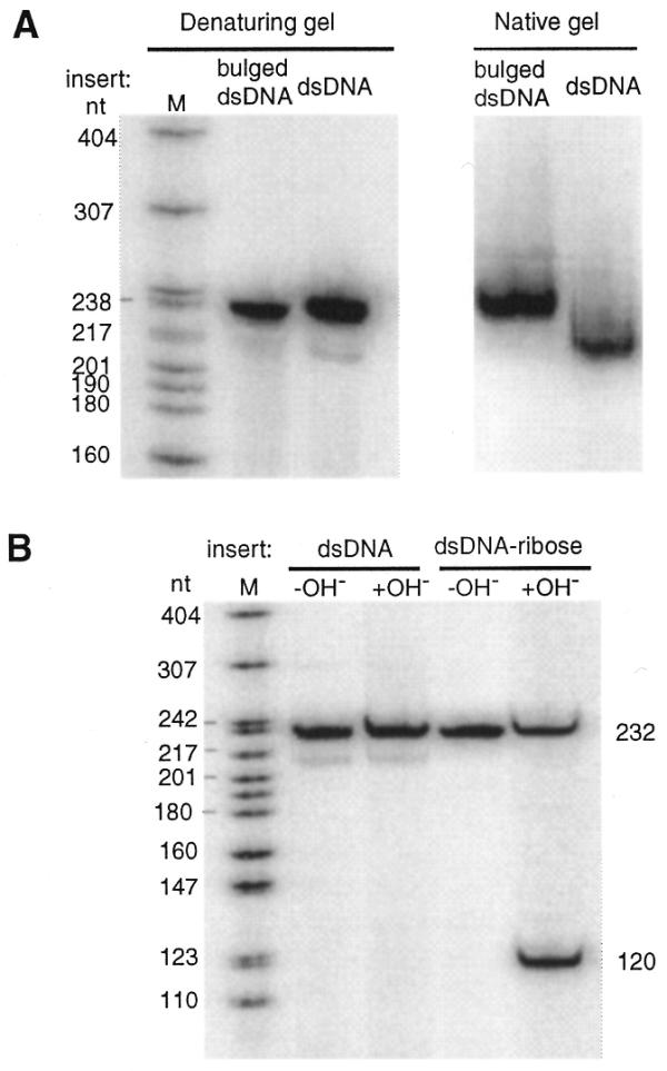
Figure 3.

Characterization of the reaction products. (A) Comparison of electrophoretic mobilities of dsDNA and bulged dsDNA. Molecular weight marker sizes (M) are shown on the left. This marker was prepared by 5′-end-labeling an MspI digest of pBR322 DNA (New England Biolabs) and boiling in formamide loading buffer prior to loading. Comparison of the electrophoretic mobilities between dsDNA and bulged dsDNA on a denaturing 10% polyacrylamide–7 M urea gel is shown on the left. The mobilities of dsDNA and bulged dsDNA show no significant differences. A 10% native gel in 0.5× TBE at 22°C is shown on the right. The mobility of bulged dsDNA is slower than dsDNA. (B) Hydrolysis analysis of a single ribose-containing chimeric dsDNA. dsDNA and the dsDNA–ribose were radiolabeled at the 5′-end. Samples were treated with (+OH–) or without (–OH–) alkali (pH 11.0) at 95°C for 30 min. Only the dsDNA–ribose sample contained a 2′-hydroxyl, as revealed by a cleavage band at the expected position.
