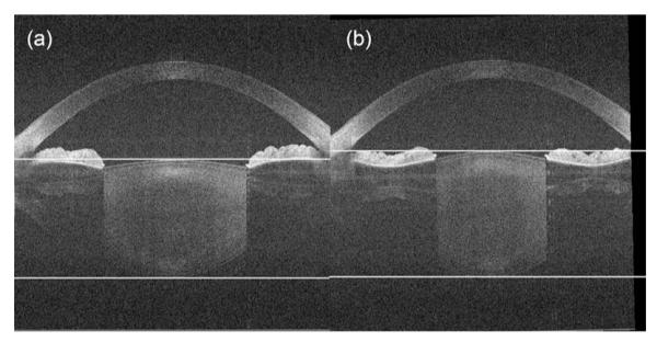
Fig. 3.

OCT B-scans during the accommodation of the eye from (a) the far point to (b) the near point. Changes of the anterior eye segment, especially the thickness of the lens, are visible. The white lines are for better visualization of the lens surface position changes during accommodation.
