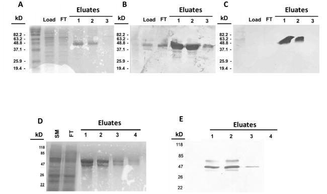
Figure 1. SDS-PAGE gels of expressed recombinant bovine PAGs-2 and 12 stained either with Coomassie blue or by Western blotting.
Panel (A) shows Coomassie blue staining of fractions from a preparation of recombinant boPAG-2. Lane-1 contains the total proteins from the lysed insect cell pellet. Lane-2 contains the flow-through of lysate from the anti-FLAG column. Lanes 3–5 contain elution fractions of recombinant boPAG-2 in order of emergence from the column. Panels (B) and (C) show Western blot images of boPAG-2-containing fractions transferred from identically loaded gels and immuno-blotted with anti-PAG-2 polyclonal and anti-FLAG monoclonal antibodies, respectively. (D) Coomassie blue staining of fractions from a preparation of recombinant boPAG-12. Lane-1 contains total proteins from the insect cell lysate. Lane-2 contains the flow-through from the anti-FLAG column. Lanes 3–5 contain fractions in order of their elution from the column. (E) Western blot image of boPAG-12-containing fractions immunoblotted and detected with anti-FLAG monoclonal antibody.

