
Figure 1.
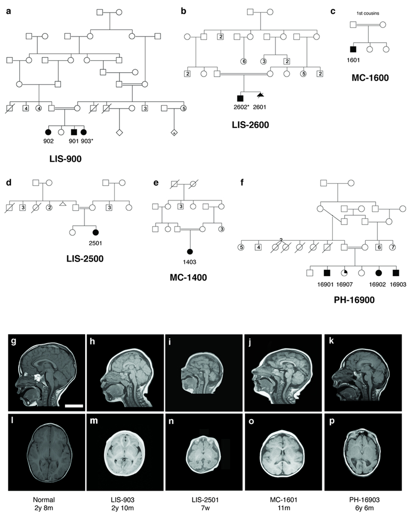
Pedigrees and radiographic findings in six consanguineous families with microcephaly and simplified gyri (MCSG). (a–f) Shaded symbols denote affected individuals. (a) LIS-900, a Mexican family with three affected children. (b) LIS-2600, a Turkish family with two affected children1. (c) LIS-2500, (d) MC-1400, and (e) MC-1600, three Turkish families (not known to be related to LIS-2600 or each other). (f) PH-16900, a Saudi family with three affected children; a fourth child (partial shading) had mild speech delay and articulation and attention difficulties but no brain abnormalities. Whole blood DNA was obtained and analyzed from each nuclear family, with the exception of LIS-2601, who died as a fetus. Asterisks denote the two individuals chosen for targeted high-throughput sequencing. (g–p) MRI features of patients with MCSG, demonstrating the breadth of cortical phenotypes associated with WDR62. Mid-sagittal T1 sections (g–k) and axial T1 sections at the level of the insula (l–p) are shown for LIS-903 (h,m), LIS-2501 (i,n), MC-1601 (j,o), and PH-16903 (k, p) individuals, as well as a control individual (g, l) age-matched to LIS-903. Common findings include microcephaly (h–k, m–p), anomalies of the corpus callosum (small splenium in h and k, absent splenium in i, thick body in k), simplification of the normal gyral pattern (h, i, k, m, n, p) as well as more variable features such as mild asymmetries of cortical size (p), possible heterotopia and cortical clefts (not shown, see Supplementary Note, Clinical Data). Scale bar = 5 cm.