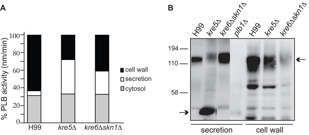
Fig. 10.
Cellular distribution of PLB activity (A) and the Plb1 protein (B). (A) Subcellular fractions were prepared from concentrated cell suspensions after an overnight incubation period and harvesting of the secreted proteins. PLB activity in the crude membrane fraction was negligible. * denotes p<0.005 (unpaired 2-tail t-test) compared to the corresponding fraction from H99. Similar results for all three strains were obtained in a second independent experiment (not shown). (B) Plb1 protein in the secreted and cell wall fractions were detected by western blotting with an anti-Plb1 peptide antibody. Top and bottom arrows indicate the position of full-length, fully glycosylated Plb1 and a breakdown product (most prominent in kre5Δ secretions), respectively.

