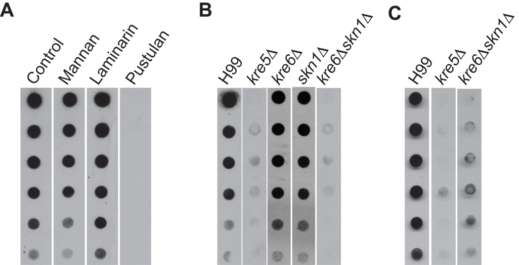
Fig. 4.
β-1,6-glucan dot blot assay. (A) Polysaccharide competition analysis. Alkali soluble cell wall material from H99 was spotted onto membrane. The primary anti-β-1,6-glucan antiserum was preincubated with the indicated purified polysaccharides for 30 min before probing the membrane. (B) Alkali-soluble cell wall β-1,6-glucan analysis of H99 and deletion strains. Cells were grown at 30°C to mid-log phase and alkali-soluble polysaccharides were isolated and spotted in 1:2 serial dilutions onto nitrocellulose membrane. Membranes were probed with polyclonal anti-β-1,6-glucan antiserum. (C) Alkali-insoluble, chitinase-released cell wall β-1,6-glucan analysis of H99 and deletion strains.

