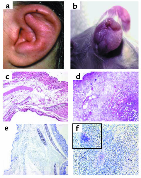
Figure 1.
Swelling and edema with a shrunken ear as observed in a human ear (a) and an affected mouse (b) with auricular chondritis. H&E-stained section of a normal (c) and a chondritic ear (d) of NOD.DQ8 transgenic mice showing massive infiltration of cells in the chondritic ear. Toludine blue staining of a normal ear (e) and a chondritic ear (f) showing destruction of cartilage in the latter, with residual cartilage seen among infiltrates as shown within the boxed area (magnification ×200). Micrographs are ×50 magnification.

