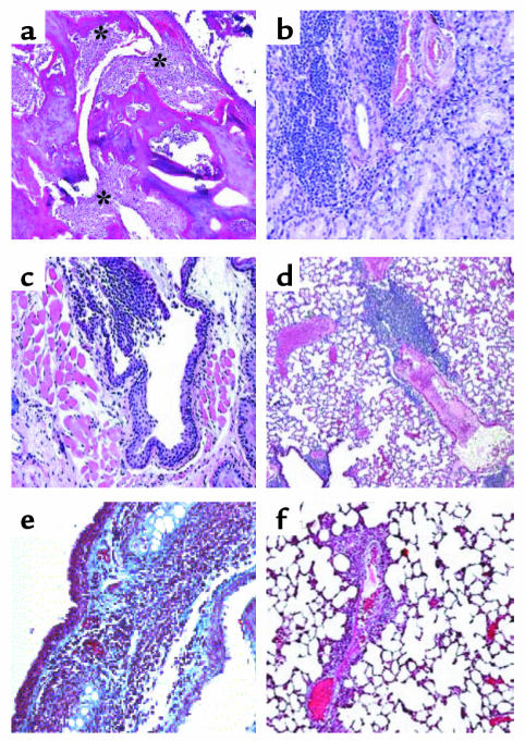
Figure 4.
Sections from various organs of NOD.DQ8 mice stained with H&E. (a) Representative inflammatory arthritis of the knee from a NOD.DQ8 mouse. Significant mononuclear infiltration with cartilage destruction and pannus formation is seen (*). (b) Salivary glands showing mononuclear infiltrate suggestive of sialadentitis. (c) Infiltration with mononuclear cells in the trachea. (d) A section of lung shows perivascular infiltration in lungs. Accustain trichrome staining of frozen sections of trachea (e) and lungs (f). Only tracheas showed deposition of collagen, as seen by the blue color. Micrographs a–d are at ×100 magnification; e and f are at ×200 magnification.

