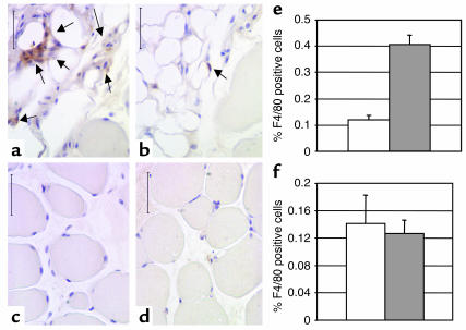
Figure 4.
Macrophages in the liver and muscle of lean and obese mice. Immunohistochemical detection of cells expressing the macrophage-specific antigen F4/80 (arrows) in extensor digitalis longus muscles from C57BL/6J (a and c) Lepob/ob female and (b and d) lean female mice. Macrophages were rarely detected in areas surrounding the myofibrils (c and d). However, muscle from both lean and obese animals was infiltrated and surrounded by adipose tissue that contained significant numbers of F4/80-positive macrophages (a and b). The percentage of F4/80-positive macrophages within this adipose tissue was markedly increased in obese compared with lean mice (e, P < 0.005). The percentage of F4/80-positive Kupffer cells within liver was not significantly altered in obesity. Calibration mark = 40 0m; white bars, lean mice; gray bars, Lepob/ob mice.

