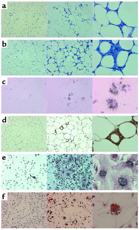
Figure 5.
Histological comparison between wild-type and ob/ob WAT and stromal-vascular cells. For each panel, the wild type at ×100 is seen at the left, ob/ob at ×100 in the middle, and ob/ob at ×400 at the right. (a) WAT morphological differences at 3 months (toluidine blue O on paraffin sections). Note the presence of nucleated stromal cells in the high magnification of the ob/ob type at the right. (b) WAT morphological differences at 5 months (toluidine blue O on paraffin sections). The stromal multinucleated cells have increased in the ob/ob type seen at the right, with early features of lipolysis in the ob/ob adipocytes manifested by multifocal cell shrinkage. (c) WAT at 3 months probed with F4/80 antisense RNA (in situ hybridization on fresh frozen sections). (d) WAT at 3 months immunostained with anti–F4/80 antibody (immunohistochemistry on paraffin sections, brown staining). (e) Primary stromal-vascular cells from 5-month-old mice, immunostained with anti–F4/80 antibody (red staining). (f) Primary stromal-vascular cells from 5-month-old mice stained with oil red O.

