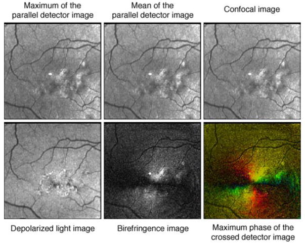
Figure 9.
Six images used in this study in a patient with non-exudative (dry) age-related macular degeneration. The fovea is not easily localizable in images that do not contain birefringence information but can easily be localized within a small area in both the Birefringence Image and the Maximum Phase of the Crossed Detector Image. The bright spot in the center of the images is a reflection artifact inherent in the GDx instrument.

